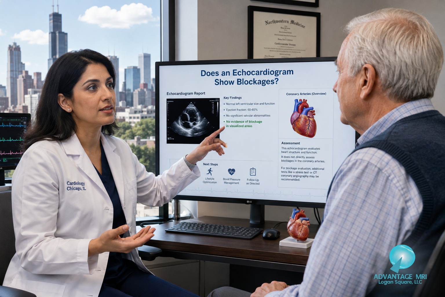
Youre sitting in a waiting room and the doctor just said the word echocardiogram. Maybe you had some chest pain or shortness of breath and now youre wondering what this test can actually find. Can it show a blockage in your arteries? Thats the question most people ask before they even walk into the room.
Heres the short answer. An echocardiogram uses sound waves to create pictures of the heart and it shows how well the heart muscle pumps blood and how the heart valves work. It does not take a direct picture of the coronary arteries the way an angiogram does. But it can pick up warning signs that point toward a blockage and thats often the first clue your doctor needs.
This article walks through what an echocardiogram actually shows, what it can’t show, and how it fits into the bigger picture of finding heart disease. For anyone wondering does an echocardiogram show blockages, it’s written for people in Chicago and across Illinois who want a plain answer without the medical jargon.
What an Echocardiogram Actually Shows
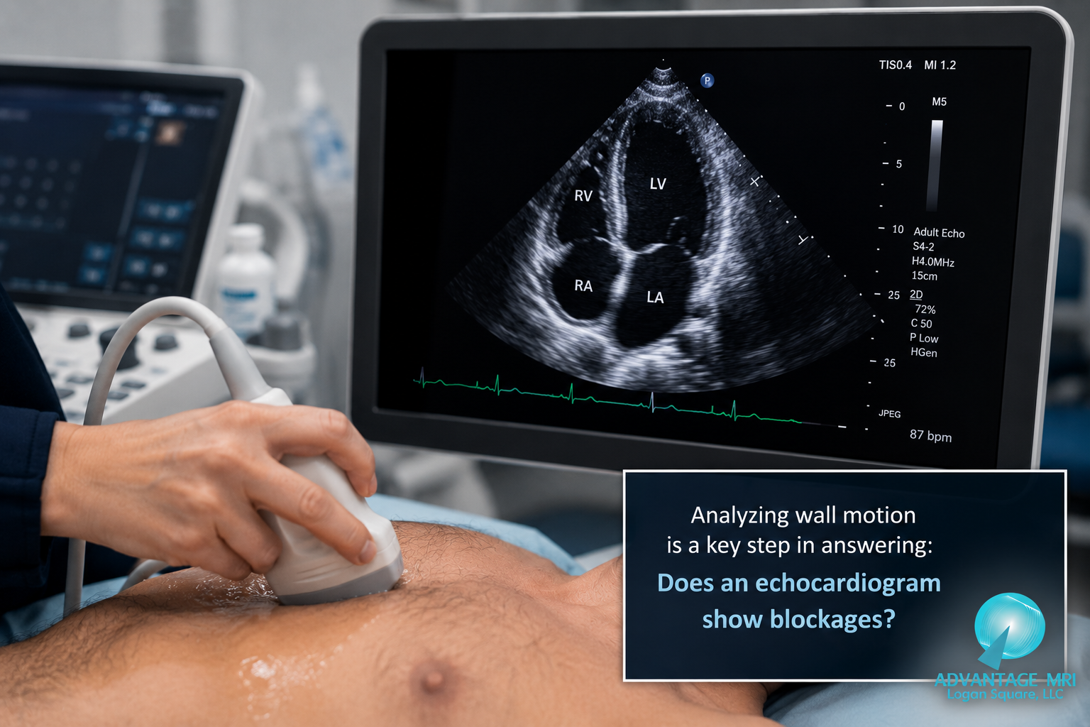
An echocardiogram is a heart ultrasound. A technician places a small device called a transducer on your chest and it sends out sound waves that bounce off the heart. Those sound waves come back and turn into moving images of your heart and blood vessels on a screen.
This test gives your doctor a detailed look at the heart chambers the heart valves and how well the heart is pumping blood with every beat. It can also show the size of the heart and whether any part of the heart muscle looks enlarged or weak.
Doctors also use echocardiograms for a lot more than checking for blockages. Patients often ask does an echocardiogram show blockages, and while doctors look into that, they also use it to help track heart failure, monitor leaky heart valves, and follow up on congenital heart defects that someone may have had since birth. A cardiologist might order one after noticing high blood pressure, heart palpitations, or unexplained shortness of breath just to get a clearer look at what’s going on before deciding on next steps. It’s also the same basic technology used for a fetal echocardiogram, which checks an unborn baby’s heart for congenital heart disease long before birth. Adult congenital heart disease clinics use the same imaging to track patients who were born with heart defects and need lifelong monitoring.
How the Ultrasound Picture Works
There is no radiation and no needles involved in a standard echocardiogram. The sound waves travel through your chest wall and bounce back at different speeds depending on what tissue they hit. A computer turns those signals into a live picture of your heart in motion. Its similar to the ultrasound used to check on an unborn babys heart just aimed at an adult heart instead.
Most people get what is called a transthoracic echocardiogram which means the probe stays on the outside of your chest. In some cases a doctor may order a transesophageal echocardiogram where a thin probe goes down the throat for a closer view. That version is less common and usually reserved for more detailed heart valve or structural questions.
What Doctors Look At on an Echocardiogram
Doctors are checking several things at once during this test. They look at how strong each heart chamber squeezes. They check whether the heart valves open and close properly or whether blood is leaking backward. They also study the overall size and shape of the heart.
| What Echo Measures | What It Shows |
|---|---|
| Heart chambers | Size and whether any chamber is enlarged |
| Ejection fraction | How much blood the heart pumps out with each beat |
| Heart valves | Whether valves open fully and close without leaking |
| Wall motion | Whether any area of heart muscle moves weaker than the rest |
| Heart rhythm | General pattern of how the heart is beating during the scan |
Can an Echocardiogram Detect a Blockage
This is where things get a little more specific. An echocardiogram can detect blockage in an indirect way. It does not look inside the artery itself. What it can show is the effect a blockage has on the heart muscle and thats often just as useful for a doctor trying to figure out whats going on.
Why Echo Doesnt See Arteries Directly
The coronary arteries are thin blood vessels that wrap around the surface of the heart and supply it with oxygen rich blood. Standard ultrasound sound waves are not designed to get a clear picture inside those small vessels. So a routine echocardiogram wont show you a picture of plaque sitting inside an artery the way a CT scan or angiogram can.
What it will show is how the heart muscle behaves when blood flow to part of it is reduced. If an area of the heart muscle isnt getting enough oxygen rich blood because of a blockage that area often moves weaker than the rest of the heart during the scan. Thats the clue a cardiologist is trained to catch.
Signs on Echo That Suggest a Blockage
A few findings on an echocardiogram can raise a flag for coronary artery disease. These include weak or abnormal wall motion in one section of the heart muscle a lower than expected ejection fraction or signs that part of the heart isnt contracting the way the rest of it is. None of these findings alone confirm a blockage but together they help a doctor decide whether more testing is needed.
Stress Echocardiogram and Coronary Artery Disease
A regular echocardiogram checks your heart at rest. A stress echocardiogram checks how your heart responds to physical activity and thats a big difference when it comes to catching blockages.
How Stress Testing Adds to a Regular Echo
During a stress echocardiogram you either walk on a treadmill or receive medication that makes your heart work harder the same way exercise would. The technician takes images of your heart before and right after that stress. If an artery is partly blocked the heart muscle it supplies may not get enough blood during exertion even if it looked fine at rest. That shows up as a new area of weak wall motion right after the stress portion of the test.
This combination test helps doctors catch blockages that a resting echo alone might miss. Its one of the more useful tools for assessing how the heart responds to physical demand without needing an invasive procedure first.
When a Doctor Might Order This Instead
Your doctor may order a stress echocardiogram if you have chest pain that happens during activity shortness of breath with exertion or risk factors for heart disease such as high blood pressure diabetes or a family history of heart disease. Its also common after a standard echo shows something worth a closer look.
Normal vs Abnormal Echocardiogram Results
Once your echocardiogram is done a cardiologist reviews the images and puts together a report. Understanding what normal and abnormal actually mean can take some of the worry out of waiting for results.
What a Normal Report Looks Like
A normal echocardiogram usually shows an ejection fraction in the typical healthy range heart valves that open and close properly with no major leaking and heart muscle that contracts evenly across all sections. A normal result is reassuring but it does not always rule out early coronary artery disease especially if the test was done at rest.
What Counts as Abnormal and Next Steps
An abnormal echocardiogram might show a lower ejection fraction leaky or narrowed heart valves an enlarged heart chamber or wall motion that looks weaker in one specific area. Depending on what the report shows your doctor may recommend more testing lifestyle changes medication or in some cases a referral to a cardiologist for further evaluation.
| Finding | Normal Result | Possible Concern |
|---|---|---|
| Ejection fraction | Around 55 to 70 percent | Below 50 percent may suggest weak pumping |
| Heart valves | Open and close smoothly | Leaking or narrowing present |
| Wall motion | Even across all areas | One area contracts weaker than others |
| Heart size | Normal chamber size | Enlarged chamber present |
This test that uses sound waves gives a strong starting point but it works best alongside your symptoms and other test results not as a stand alone diagnosis.
Echocardiogram vs Other Heart Tests for Blockages
No single test tells the whole story about your heart. Doctors often combine an echocardiogram with other tools depending on your symptoms and risk factors.
Echo vs EKG
An EKG testing session records the electrical activity of your heart and can catch rhythm problems or signs of a past heart attack in seconds. An echocardiogram instead shows the physical structure and pumping function of the heart. Many doctors order both because they answer different questions. One looks at electrical signals and the other looks at motion and structure.
Echo vs CT Angiogram
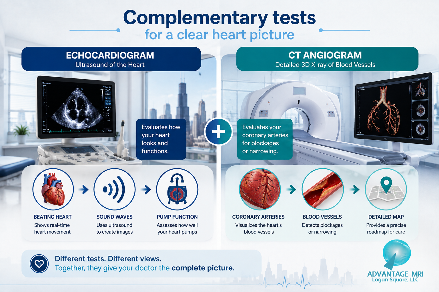
A CT scan angiogram uses contrast dye and imaging to create a direct picture of the coronary arteries themselves showing exactly where a blockage sits and how severe it is. This is closer to a direct blockage test than an echocardiogram. Advanced MRI imaging can also provide detailed pictures of the heart and blood vessels when a doctor needs a closer structural look beyond what ultrasound alone can offer.
When a Doctor Adds Other Tests for Related Symptoms
Sometimes chest discomfort or numbness in the arms and legs isnt about the heart at all. A doctor might order EMG testing or a nerve conduction study to rule out nerve related causes of pain or tingling that can sometimes feel similar to heart symptoms. Its a good reminder that heart tests and nerve tests both play a role in figuring out whats really going on.
What Happens If Your Echocardiogram Shows a Problem
Getting an abnormal result can feel scary. But an abnormal echocardiogram is a starting point for answers not a final diagnosis on its own.
Follow Up Testing Your Doctor May Order
If your echo shows a possible blockage your doctor might order a stress test a CT angiogram or refer you for a cardiac catheterization to look directly inside the arteries. In some cases a holter monitor may be used for a few days to track your heart rhythm continuously and catch anything an echo cant.
Getting a Second Opinion or a Confirming Scan
If results are unclear or borderline asking for a repeat scan or a second opinion is a completely normal step. Heart health decisions shouldnt feel rushed and a good imaging center will walk you through what your results actually mean before recommending next steps.
Getting an Echocardiogram in Chicago
If youre in the Hermosa neighborhood or anywhere around Chicago and your doctor has recommended an echocardiogram Advantage MRI offers this test along with a full range of diagnostic imaging services in one location.
What to Expect at Your Appointment
The test itself is painless and usually takes under an hour. Youll lie back while a technician moves the ultrasound probe across your chest to capture images from different angles. There is no recovery time needed afterward and you can drive yourself home. If your doctor also wants imaging like a digital x ray or a heart ultrasound as part of your workup those can often be scheduled together to save you a trip.
Why Choose an ACR Accredited Center
Advantage MRI is ACR accredited which means the equipment training and image quality meet standards set by the American College of Radiology. Reports are reviewed by board certified radiologists which supports accurate reads and faster turnaround for your referring doctor. You can learn more about us and our approach to imaging care.
Dr Gregory T Goldstein MD leads the imaging program at Advantage MRI. Hes a board certified diagnostic radiologist with a background in orthopedic and neurological imaging and hes also the founder of MetisMD and the creator of Connected RadiologySM a model built around faster reporting and closer coordination with referring physicians. That kind of oversight matters when the images being reviewed could shape decisions about your heart health and reduce the guesswork for your doctor.
Getting scheduled is usually simple too. Most patients can get an appointment within a few days and results go back to your referring physician quickly so you arent left waiting weeks to find out whether your visit turned up anything worth a follow up conversation.
Frequently Asked Questions
Does an echocardiogram show blockages in arteries?
Not directly. It shows how the heart muscle moves and pumps and weak areas of movement can point toward a blockage even though the test doesnt picture the artery itself.
Can an echocardiogram detect blocked heart arteries directly?
No it cant see inside the arteries the way a CT angiogram or cardiac catheterization can. It picks up the downstream effects of a blockage on heart muscle function instead.
What can an echocardiogram show about heart health?
It shows heart chamber size ejection fraction valve function and how evenly the heart muscle contracts. Its a broad snapshot of how well your heart is working overall.
Whats the difference between normal and abnormal echocardiogram results?
A normal result shows even wall motion a healthy ejection fraction and valves working properly. An abnormal result shows a weak area of muscle movement a lower ejection fraction or valve problems that need follow up.
Is a stress echocardiogram better for finding blockages?
Its more sensitive than a resting echo because it shows how the heart performs under exertion which is often when a partial blockage causes a problem.
How long does an echocardiogram take?
Most exams take between thirty and sixty minutes depending on how many views the technician needs to capture.
Does an echocardiogram hurt or use radiation?
No. It uses sound waves not radiation and most people feel nothing beyond mild pressure from the probe against the chest.
What test finds an artery blockage if echo cant confirm it?
A CT angiogram or cardiac catheterization gives a direct look inside the coronary arteries and is typically used to confirm what an echo suggests.
Can you have coronary artery disease with a normal echo?
Yes especially early on. A resting echo can look normal even when a partial blockage exists which is why a stress test or additional imaging sometimes gets added.
How soon do I get echocardiogram results?
Many centers including Advantage MRI aim for fast turnaround so your referring doctor gets a report quickly and can discuss next steps with you sooner rather than later.
Conclusion
An echocardiogram is one of the most useful heart tests out there and it tells your doctor a lot about how your heart is functioning. It doesnt take a direct picture of a blockage inside your coronary arteries but it often shows the warning signs a blockage leaves behind. Combined with a stress test an EKG or a CT angiogram when needed it becomes part of a bigger picture that helps your doctor keep your heart health on track.
This article is for general information only and is not a substitute for medical advice diagnosis or treatment from your doctor. Always talk with a qualified healthcare provider about your specific symptoms and test results.
Contact Us
- Address: 2008 North Pulaski Road Chicago IL 60639
- Phone: (773) 227 9777
- Contact form: available on our contact us page for general questions
For reliable advanced diagnostic imaging you can trust call Advantage MRI today or schedule your appointment online.
Follow us on social media for the latest health tips diagnostic imaging updates and clinic news: Facebook | Instagram | LinkedIn.