Health & Imaging
What Is an Echocardiogram? Types, Procedure and What Your Results Mean

What is an echocardiogram? Quick answer: Often called an “echo,” it is an ultrasound of the heart that uses high-frequency sound waves, not radiation, to create real-time moving pictures of your heart. An echo shows the size and shape of your heart’s chambers, how thick the walls are, how well the heart pumps (ejection fraction), how the four valves open and close, and how blood flows through the heart. It can also detect fluid around the heart, signs of high pressure in the lungs, and certain congenital defects. The most common type, a transthoracic echocardiogram (TTE), is done by moving a probe over your chest, is painless, and usually takes 30 to 60 minutes.
This pillar guide explains what an echocardiogram is, what it shows and doesn’t show, the different types, why doctors order it, how to prepare, what happens during the test, what your results mean, and what usually comes next.
Need to Schedule a Diagnostic Appointment?
Whether you need an MRI, ultrasound, X-ray, or another diagnostic service, Advantage MRI is here to help you take the next step in your healthcare journey.
Submit your appointment request online, and our team will contact you to confirm your visit.
Book Your Appointment →How Does an Echocardiogram Work?
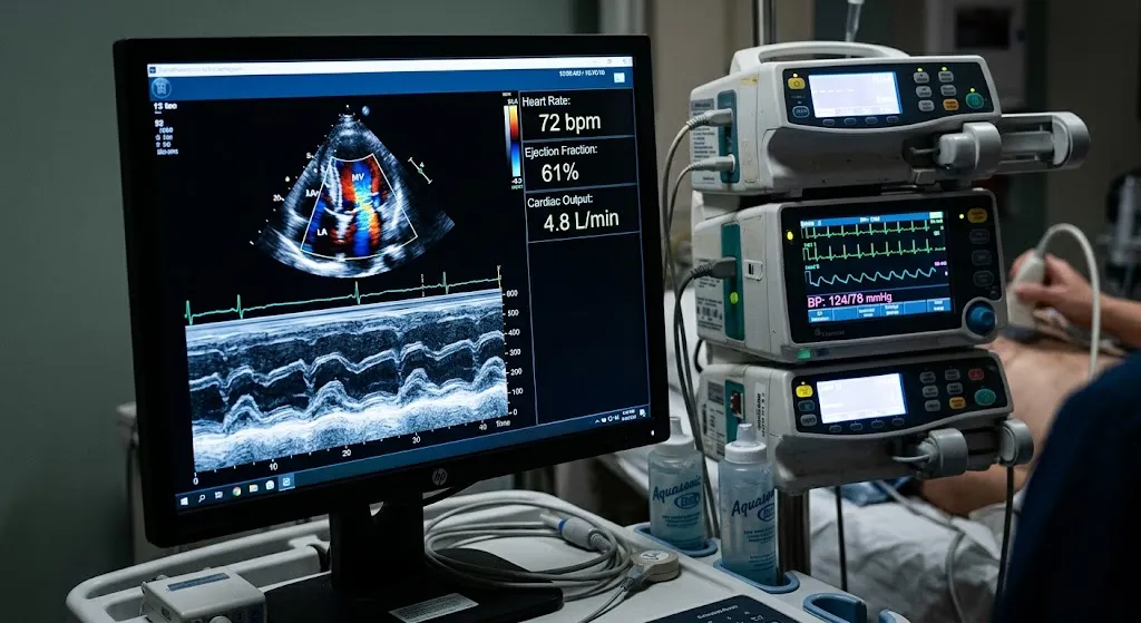
An echocardiogram works like other ultrasound exams. A handheld probe (transducer) sends sound waves into your chest. The waves bounce off the heart’s structures and return to the probe, and a computer turns those echoes into moving images. Because the images are in real time, doctors can watch your heart beat, see the valves move and measure how the walls contract.
Doppler ultrasound, built into the echo machine, measures the speed and direction of blood flow. It’s shown as color maps and waveforms and helps detect leaky or narrowed valves, holes between chambers, and pressures inside the heart.
What Does an Echocardiogram Show?
Heart Pumping Function (Ejection Fraction)
Ejection fraction (EF) is the percentage of blood the left ventricle, the heart’s main pumping chamber, pushes out with each beat. It’s one of the most important numbers on an echo report.
| Ejection fraction | General interpretation |
|---|---|
| About 50-70% | Generally considered normal |
| 41-49% | Mildly reduced |
| 40% or lower | Reduced; may indicate heart failure with reduced ejection fraction |
| Above about 70% | Higher than usual; sometimes seen with certain conditions |
Ranges vary slightly by laboratory and guideline, and a normal EF doesn’t rule out all heart problems. Some people have heart failure with a preserved (normal) EF.
Chamber Size
An echo measures the size of the four chambers: left and right atria (upper chambers) and left and right ventricles (lower chambers). Enlarged chambers can be a sign of conditions such as valve disease, cardiomyopathy or long-standing high blood pressure.
Wall Thickness
Thickened heart walls (hypertrophy) can result from high blood pressure, aortic valve narrowing, or inherited conditions such as hypertrophic cardiomyopathy.
Wall Motion
The echo shows whether all parts of the heart wall contract normally. An area that doesn’t move well may suggest damage from a past heart attack or reduced blood flow.
Heart Valves
Echocardiography is the main test for valve problems:
- Stenosis: a valve that’s narrowed and doesn’t open fully
- Regurgitation: a valve that leaks backward
- Prolapse: a valve leaflet that bulges backward, such as mitral valve prolapse
Pressures and Blood Flow
Doppler measurements can estimate pressures inside the heart and in the lungs (pulmonary artery pressure), which helps identify pulmonary hypertension.
Fluid Around the Heart
An echo can detect pericardial effusion, a buildup of fluid in the sac around the heart.
Congenital Heart Defects
Holes between chambers, such as an atrial septal defect, and other structural abnormalities present from birth can be identified.
Clots and Masses
An echo can sometimes show blood clots or tumors inside the heart. A transesophageal echo is more sensitive for some of these.
What an Echocardiogram Doesn’t Show
- Coronary arteries directly. A standard echo can’t see the inside of the arteries that supply the heart muscle. Indirect signs of blockage, like abnormal wall motion, may appear, especially during a stress echo. See can an echocardiogram detect a blocked artery?
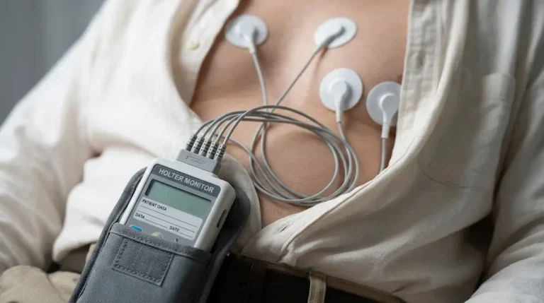
- Heart rhythm over time. An echo shows rhythm only during the test. Holter or event monitors are used for intermittent rhythm problems. See What Is a Holter Monitor?
- Every structure clearly in every person. Body size, lung disease and chest anatomy can limit image quality.
Why Would a Doctor Order an Echocardiogram?
Common reasons include:
- A heart murmur heard during an exam
- Shortness of breath, especially with exertion or lying flat
- Swelling in the legs or ankles
- Chest pain, sometimes as part of a stress echo
- Palpitations or an abnormal EKG
- Fainting or dizziness
- High blood pressure, to check for heart muscle thickening
- Suspected heart failure or monitoring known heart failure
- After a heart attack, to assess damage and pumping function
- Valve disease monitoring or before and after valve procedures
- Atrial fibrillation, to check heart structure
- Stroke, to look for a source of clots
- Certain cancer treatments that can affect heart function
- Pregnancy-related concerns or a family history of inherited heart conditions
Types of Echocardiograms
| Type | How it’s done | Common uses |
|---|---|---|
| Transthoracic echo (TTE) | Probe moved over the chest; most common | General evaluation of structure, function and valves |
| Transesophageal echo (TEE) | Thin probe passed down the throat under sedation | Detailed valve views, clots in the heart, infections on valves, before some procedures |
| Stress echocardiogram | Images before and after exercise or medication | Evaluating reduced blood flow from coronary artery disease |
| Doppler echocardiogram | Part of most echos; measures blood flow | Valve leaks and narrowing, pressures |
| Contrast (enhanced) echo | IV microbubble agent improves images | When standard images are unclear |
| 3D echocardiography | Three-dimensional images | Detailed valve and chamber assessment |
| Fetal echocardiogram | Done during pregnancy on the baby’s heart | Detecting congenital heart defects before birth |
To compare the two main approaches, read TTE vs TEE: transthoracic and transesophageal echocardiograms compared. For stress testing, see stress echocardiogram vs stress EKG.
How to Prepare for an Echocardiogram
Transthoracic Echo (Most Common)
- Eat, drink and take medications normally, unless told otherwise.
- Wear a two-piece outfit, since you’ll undress from the waist up.
- Avoid lotion or oil on your chest.
- Bring your ID, insurance card, doctor’s order and medication list.
Transesophageal Echo
- Fast for several hours beforehand, as instructed.
- Arrange a ride home, because sedation is used.
- Tell the team about swallowing problems, esophageal conditions or medication allergies.
Stress Echo
- Follow instructions about eating, caffeine and certain heart medications.
- Wear comfortable walking shoes and clothing if you’ll exercise.
For more on preparation for heart and imaging tests, see how to prepare for an MRI, echocardiogram or ultrasound.
What Happens During a Transthoracic Echocardiogram?
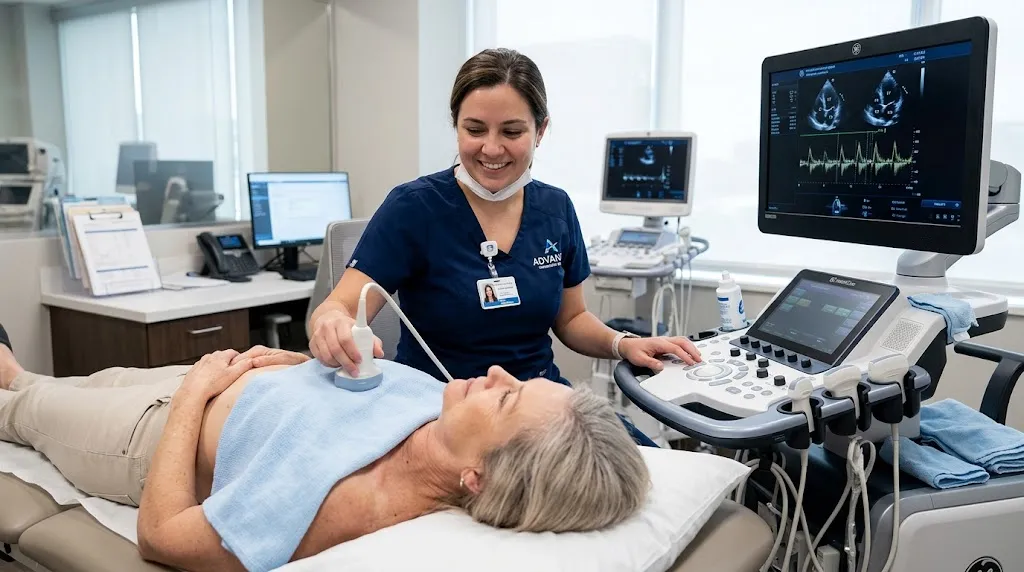
- Changing: You’ll remove clothing from the waist up and wear a gown that opens in the front.
- Electrodes: Three small electrodes are placed on your chest to track your heart rhythm during imaging.
- Positioning: You’ll lie on your left side on an exam table, often with your left arm raised.
- Gel and probe: Gel is applied, and the sonographer presses the probe against your chest at several locations: beside the breastbone, near the tip of the heart (often under the left breast), below the ribs and above the collarbone.
- Breathing instructions: You may be asked to hold your breath briefly or change position.
- Doppler sounds: You may hear whooshing sounds from blood flow.
- Contrast (if needed): A microbubble agent may be given through an IV to improve images.
- Finishing: The gel is wiped off, and you can return to normal activities right away.
Women often have questions about undressing and positioning. We cover that in detail in how is an echocardiogram done on a woman?
How Long Does an Echocardiogram Take?
A standard transthoracic echo usually takes 30 to 60 minutes. A TEE takes longer because of sedation and recovery, and a stress echo often takes about an hour or more. See how long does an echocardiogram take? for a full timeline.
Is an Echocardiogram Safe?
Yes. A transthoracic echo uses sound waves, not radiation, and has no known harmful effects. You might feel firm pressure from the probe. A TEE carries small risks related to sedation and passing the probe, such as a sore throat. Stress echos involve monitored exercise or medication. Contrast agents are generally well tolerated; tell the team about allergies.
Echocardiogram vs EKG
These two tests are often ordered together, but they measure different things:
| Echocardiogram | EKG | |
|---|---|---|
| What it measures | Structure and function (pictures) | Electrical activity (wave tracing) |
| Technology | Ultrasound | Electrodes recording signals |
| Time | 30-60 minutes | About 5-10 minutes |
| Shows | Pumping, valves, chambers, walls | Rate, rhythm, conduction, heart attack signs |
Learn more in echocardiogram vs EKG and What Is an EKG Test?
Conditions an Echocardiogram Helps Diagnose or Monitor
| Condition | What the echo typically shows |
|---|---|
| Heart failure | Reduced or preserved ejection fraction, enlarged chambers, abnormal filling |
| Cardiomyopathy | Weakened, enlarged or abnormally thickened heart muscle |
| Aortic stenosis | Narrowed aortic valve with high flow speeds on Doppler |
| Mitral regurgitation | Backward leak through the mitral valve |
| Pulmonary hypertension | Elevated estimated lung pressures and right-heart strain |
| Pericarditis or pericardial effusion | Fluid or thickening around the heart |
| Congenital heart disease | Holes or structural abnormalities present from birth |
| Endocarditis | Infection on a valve (TEE is often needed for detail) |
| After a heart attack | Areas of the wall that don’t move normally; overall pumping |
Echocardiograms in Specific Situations
During Pregnancy
Echocardiograms are safe in pregnancy because ultrasound uses no radiation. They’re used when a pregnant person has a murmur, shortness of breath beyond what’s expected, swelling, palpitations or a known heart condition. Pregnancy normally increases blood volume and heart workload, and the reader takes that into account.
Children
Pediatric echocardiograms evaluate murmurs, congenital heart defects and symptoms such as fainting or chest pain. They’re usually interpreted by pediatric cardiologists, and young children may need comfort measures or distraction to stay still.
Athletes
Endurance athletes can develop a larger, thicker “athlete’s heart,” which is a normal adaptation. An echo helps distinguish this from inherited conditions that need different management.
How Often Do You Need an Echocardiogram?
There’s no routine schedule for healthy people. Repeat echos are usually based on findings and guidelines. For example, someone with a mild leaky valve might be rechecked every few years, while someone with a moderate or severe valve problem, or with heart failure being treated with medication, might be rechecked more often. Your doctor sets the interval. Keeping copies of earlier reports, or having them done at the same facility, makes comparison easier.
Understanding Your Echocardiogram Results
A cardiologist or qualified physician interprets your echo and sends a report to your doctor. Common terms include:
| Term | What it means |
|---|---|
| LVEF (left ventricular ejection fraction) | Percentage of blood pumped out of the main chamber each beat |
| Normal LV size and function | Main pumping chamber is normal in size and pumps well |
| LVH (left ventricular hypertrophy) | Thickened walls of the main pumping chamber |
| Diastolic dysfunction | The heart muscle is stiffer and doesn’t relax normally between beats; common with age and high blood pressure |
| Trace or mild regurgitation | A small valve leak; trace leaks are common and often normal |
| Moderate or severe regurgitation | A larger leak that may need monitoring or treatment |
| Stenosis | Narrowing of a valve |
| Regional wall motion abnormality | Part of the heart wall doesn’t move normally |
| Pericardial effusion | Fluid around the heart |
| RVSP / PASP | Estimated pressure in the lungs’ arteries |
Seeing words like “mild” or “trace” is common and often not a concern. Always review results with your doctor.
What Happens After an Echocardiogram?
Depending on your results, your doctor may:
- Reassure you that your heart structure and function are normal
- Repeat the echo in months or years to monitor a valve or the heart’s pumping
- Adjust or start medications, such as for blood pressure or heart failure
- Order further tests, such as a stress test, Holter monitor, cardiac MRI or catheterization
- Refer you to a cardiologist or cardiac surgeon for valve or structural treatment
How Much Does an Echocardiogram Cost?
Cost depends on the type of echo, whether contrast is used, your insurance, the facility and whether the interpretation is billed separately. Independent outpatient centers often charge less than hospital-based departments. Billing uses specific CPT codes for each type of echo; see echocardiogram CPT codes explained. Ask for an estimate before your appointment if you’re paying out of pocket.
Echocardiograms at Advantage MRI in Chicago
Advantage MRI performs echocardiograms at 2008 N Pulaski Rd, Chicago, IL 60639. Because we also offer EKGs, Holter monitoring, ultrasound, MRI, X-ray, EMG and nerve conduction studies, patients who need more than one test may be able to complete them in one location. Results are sent to your referring physician.
Our website emphasizes a comfortable, patient-first experience. If you’d like a chaperone or have other preferences, let us know when you schedule. Learn more on our About page. We welcome insured, out-of-network and self-pay patients.
Frequently Asked Questions
What is an echocardiogram test?
It’s an ultrasound of the heart that uses sound waves to create moving images of the heart’s chambers, walls and valves and to measure blood flow.
What does an echocardiogram show?
It shows heart size, wall thickness, pumping strength (ejection fraction), valve function, blood flow, pressure estimates, fluid around the heart, and some congenital defects.
Is an echocardiogram painful?
A standard transthoracic echo isn’t painful. You may feel firm pressure from the probe and coolness from the gel.
How long does an echocardiogram take?
Usually 30 to 60 minutes for a standard transthoracic echo.
Can an echocardiogram detect a heart attack?
It can show areas of the heart wall that aren’t moving normally, which may indicate damage from a heart attack. Diagnosis of an acute heart attack also relies on symptoms, EKG and blood tests.
Can an echocardiogram detect blocked arteries?
Not directly. A stress echo can show indirect signs of reduced blood flow. Other tests are used to see the arteries themselves.
Do I need to fast before an echocardiogram?
Not for a standard transthoracic echo. Fasting is usually required for a transesophageal echo.
What is a normal ejection fraction?
Roughly 50 to 70 percent is generally considered normal, though ranges vary slightly by laboratory.
Is an echocardiogram the same as an EKG?
No. An echocardiogram is an ultrasound that shows the heart’s structure and function. An EKG records electrical activity.
Conclusion
An echocardiogram is one of the most valuable tools in heart care. In about half an hour to an hour, without radiation or pain, it shows how your heart is built and how it works: chamber size, wall thickness, pumping strength, valve function and blood flow. Different types, from standard transthoracic echos to TEE and stress echos, answer different questions. Understanding what an echo shows, how to prepare and what your results mean helps you take an active role in your care.
Need an echocardiogram in Chicago? Book an appointment with Advantage MRI or call (773) 227-9777. Hours: Monday-Friday 9:00 AM-5:00 PM, Saturday 9:00 AM-1:00 PM. Follow us on Facebook and Instagram.