Medical Imaging
What Does a Brain MRI Show?
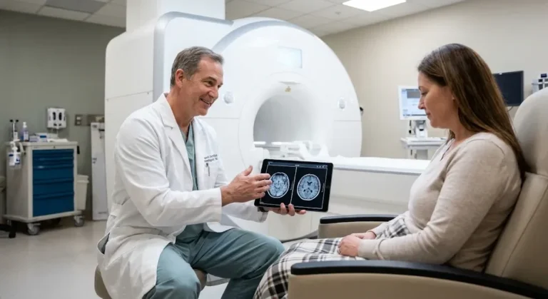
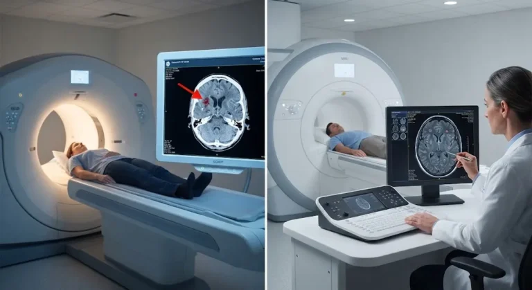
Brain MRI what does it show? A brain MRI uses magnetic fields and radio waves to create detailed images of the brain, detecting conditions such as tumors, strokes, multiple sclerosis, aneurysms and structural abnormalities. It provides superior soft tissue detail compared to CT scans without exposing patients to ionizing radiation.
Why Your Doctor May Order a Brain MRI
Brain MRI is one of the most commonly ordered neurologic imaging studies. Physicians request brain MRI when they need to visualize the brain’s structures in detail to diagnose or rule out specific conditions. Common reasons include:
Persistent or Severe Headaches
While most headaches do not require imaging, your doctor may order a brain MRI if you experience:
Need to Schedule a Diagnostic Appointment?
Whether you need an MRI, ultrasound, X-ray, or another diagnostic service, Advantage MRI is here to help you take the next step in your healthcare journey.
Submit your appointment request online, and our team will contact you to confirm your visit.
Book Your Appointment →- Sudden, severe headaches unlike any you have had before (thunderclap headache).
- Headaches that progressively worsen over weeks or months.
- Headaches accompanied by neurological symptoms like vision changes, weakness or confusion.
- New headache patterns in patients over 50.
- Headaches that wake you from sleep.
- Headaches that do not respond to standard treatment.
A brain MRI for headache evaluation can identify or exclude conditions such as tumors, aneurysms, sinus disease, Chiari malformations and increased intracranial pressure.
Seizures
New-onset seizures in adults typically require brain MRI to identify potential structural causes including tumors, vascular malformations, areas of cortical scarring or developmental abnormalities. MRI helps neurologists determine whether seizures have a treatable structural source.
Stroke Symptoms
When a patient presents with sudden weakness, numbness, speech difficulty, vision loss or coordination problems, brain imaging is essential. While CT is often the first imaging study performed in emergency settings because of its speed, MRI provides more detailed information about:
- The location and extent of stroke damage.
- Whether the stroke is ischemic (caused by a blood clot) or hemorrhagic (caused by bleeding).
- How much brain tissue can potentially be saved with treatment.
- Evidence of prior small strokes that may not have caused noticeable symptoms.
Diffusion-weighted imaging (DWI), a specialized MRI sequence, can detect ischemic stroke within minutes of onset — earlier than CT in many cases.
Suspected Brain Tumors
Brain MRI is the primary imaging tool for detecting and characterizing brain tumors. It can reveal:
- The tumor’s exact location and size.
- Whether the tumor appears benign or has features suggesting malignancy.
- The tumor’s relationship to critical brain structures.
- Associated swelling (edema) in surrounding brain tissue.
- Multiple tumors, which may suggest metastatic disease (cancer that has spread from elsewhere).
Contrast-enhanced brain MRI using gadolinium is almost always required for thorough tumor evaluation. The contrast agent highlights areas where the blood-brain barrier is disrupted, which is a common feature of many brain tumors.
Multiple Sclerosis (MS)
MRI is the most important diagnostic tool for multiple sclerosis. MS causes characteristic lesions (plaques) in the white matter of the brain and spinal cord. Brain MRI can:
- Detect MS lesions and show their distribution pattern.
- Distinguish active (enhancing) lesions from chronic ones using contrast.
- Monitor disease progression over time.
- Help differentiate MS from other conditions that produce similar symptoms.
Neurologists typically follow specific MRI protocols for MS that include contrast-enhanced sequences and may also image the spinal cord.
Cognitive Decline and Memory Loss
When patients present with progressive memory problems, confusion or behavioral changes, brain MRI helps evaluate for:
- Alzheimer’s disease and other forms of dementia (showing patterns of brain atrophy).
- Normal pressure hydrocephalus (enlarged ventricles with normal cerebrospinal fluid pressure).
- Chronic subdural hematomas.
- Brain tumors that may present with cognitive changes rather than typical neurological symptoms.
- Vascular dementia caused by multiple small strokes.
Other Reasons for Brain MRI
Additional indications include:
- Dizziness and balance problems: Evaluating the inner ear structures and brainstem.
- Vision or hearing changes: Assessing optic nerves, visual pathways and auditory nerves.
- Pituitary disorders: Identifying pituitary tumors that can affect hormone production.
- Developmental abnormalities in children: Evaluating brain structure in children with developmental delays.
- Pre-surgical planning: Mapping brain structures before neurosurgery.
- Trauma follow-up: Evaluating persistent symptoms after head injury when CT was initially normal.
What Can a Brain MRI Detect?
Brain MRI can identify a wide range of conditions affecting the brain and surrounding structures:
Tumors and Masses
Both primary brain tumors (originating in the brain) and metastatic tumors (spreading from other body areas) appear on MRI. Common types include gliomas, meningiomas, pituitary adenomas and schwannomas. Contrast enhancement helps characterize tumors and guide biopsy or surgical planning.
Stroke and Vascular Disease
MRI detects both acute and chronic strokes, cavernous malformations, arteriovenous malformations (AVMs) and small vessel ischemic disease. MR angiography (MRA) can evaluate blood vessels without invasive catheter-based procedures.
Infections and Abscesses
Brain infections including abscesses, encephalitis and meningitis produce characteristic patterns on MRI. Diffusion-weighted imaging is particularly helpful for identifying brain abscesses.
Demyelinating Diseases
Multiple sclerosis and other conditions that damage the myelin sheath surrounding nerve fibers show distinct patterns on brain MRI. These are often best visualized on FLAIR (fluid-attenuated inversion recovery) sequences.
Congenital and Structural Abnormalities
Chiari malformations, arachnoid cysts, cortical malformations and other developmental variants are clearly seen on MRI.
Hydrocephalus
Enlarged ventricles indicating excess cerebrospinal fluid accumulation are easily identified and monitored with MRI.
Aneurysms
MR angiography can detect cerebral aneurysms — weak spots in blood vessel walls that can potentially rupture and cause life-threatening bleeding.
Brain MRI vs CT Scan: Which Is Better?
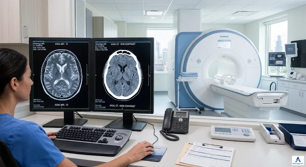
Both MRI and CT scan have roles in brain imaging. The choice depends on the clinical situation.
Brain MRI vs Brain CT Scan Comparison
| Feature | Brain MRI | Brain CT Scan |
| Radiation | No ionizing radiation | Uses ionizing radiation (X-rays) |
| Soft Tissue Detail | Superior — best for brain tissue imaging | Good but less detailed for soft tissues |
| Bone Imaging | Limited bone detail | Excellent for skull fractures and bone lesions |
| Scan Time | 30-60 minutes | 5-10 minutes |
| Emergency Use | Used for specific emergency protocols (stroke DWI) | Preferred first-line imaging in most emergencies |
| Tumor Detection | Superior sensitivity and characterization | Can detect large tumors but limited characterization |
| MS Detection | Essential diagnostic tool | Poor sensitivity for MS lesions |
| Acute Bleeding | Detectable but CT is faster for initial evaluation | Excellent for acute hemorrhage detection |
| Claustrophobia | Can be challenging (open MRI option available) | Less confining, faster scan |
| Cost | Generally higher | Generally lower |
| Contrast Agent | Gadolinium (IV) | Iodine-based (IV) |
In general, MRI provides significantly better detail for evaluating brain soft tissue, making it the preferred study for most non-emergency neurological conditions. CT excels in emergency settings where speed is critical, such as initial evaluation of head trauma or suspected acute hemorrhagic stroke.
What to Expect During a Brain MRI
Before the Scan
You will complete a safety screening questionnaire and remove all metal objects. If contrast is ordered, an IV line will be placed. The technologist will explain the procedure and provide earplugs or headphones.
During the Scan
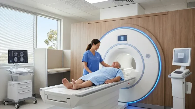
You will lie on your back on the MRI table. A head coil (a device shaped like a cage or helmet) is placed around your head to capture the magnetic signals. The table slides into the scanner.
Brain MRI scans typically take 30 to 45 minutes without contrast and 45 to 60 minutes with contrast. During the scan:
- Remain as still as possible. Even small movements can affect image quality.
- You will hear loud knocking, buzzing and clicking sounds.
- The technologist communicates through an intercom and monitors you at all times.
- You will have a call button to alert staff if needed.
- If contrast is used, it is injected partway through the scan.
After the Scan
You can resume normal activities immediately. If you received contrast, drinking extra water helps your kidneys flush the gadolinium. Your images are reviewed by a board-certified radiologist who creates a detailed report for your referring physician.
How to Prepare for a Brain MRI
Preparation for brain MRI is straightforward:
- Remove all metal jewelry, hair clips, piercings and accessories.
- Wear comfortable clothing without metal fasteners.
- Inform the imaging center about metal implants, pacemakers or other medical devices.
- Follow fasting instructions if contrast is ordered (typically 4 to 6 hours).
- Continue regular medications unless your doctor advises otherwise.
- Tell your doctor about claustrophobia before the appointment so accommodations can be arranged.
- Bring your referral, insurance card and photo ID.
If you experience significant claustrophobia, discuss open MRI options or sedation with your doctor before the appointment.
How Long Does It Take to Get Brain MRI Results?
After your brain MRI, a board-certified radiologist reviews the images and creates a diagnostic report. This report is sent to your referring physician, who will discuss the findings with you.
At Advantage MRI in Chicago, efficient reporting ensures your doctor receives results as quickly as possible. Turnaround times vary depending on the complexity of the study, but most reports are available within 24 to 48 hours.
If your brain MRI reveals urgent findings, the radiologist may contact your physician immediately so treatment can begin without delay.
Brain MRI at Advantage MRI in Chicago
Advantage MRI at 2008 North Pulaski Road, Chicago, IL 60639 provides comprehensive brain MRI services including:
- Standard and contrast-enhanced brain MRI.
- MR angiography (MRA) for blood vessel evaluation.
- Open MRI for patients with claustrophobia.
- ACR-accredited imaging equipment meeting rigorous quality standards.
- Board-certified radiologists with neurologic imaging expertise.
- Efficient reporting for timely results.
To book a brain MRI in Chicago or ask questions about your upcoming study, call Advantage MRI at (773) 227-9777.
Conclusion
Brain MRI is a powerful and safe diagnostic tool that provides unmatched detail of brain structures without radiation exposure. Whether your doctor is investigating headaches, stroke symptoms, suspected tumors or neurological conditions like multiple sclerosis, brain MRI delivers the information needed for accurate diagnosis and treatment planning.
If your physician has recommended a brain MRI, Advantage MRI in Chicago offers ACR-accredited imaging with board-certified radiologists. Call (773) 227-9777 to schedule your appointment or learn more about the brain MRI process. Follow us on Facebook and Instagram. Learn more about a Chicago echocardiogram at Advantage MRI.
Frequently Asked Questions About Brain Mri What Does It Show
1. What does a brain MRI show?
A brain MRI shows detailed images of brain tissue, blood vessels, nerves and surrounding structures. It can detect tumors, strokes, multiple sclerosis, aneurysms, infections, structural abnormalities and signs of dementia.
2. Is a brain MRI painful?
No. Brain MRI is completely painless. You may hear loud noises from the scanner and feel the table vibrate slightly, but the exam itself does not cause discomfort. If contrast is used, you may feel a brief cool sensation from the IV injection.
3. How long does a brain MRI take?
A standard brain MRI without contrast takes approximately 30 to 45 minutes. With contrast, the scan typically takes 45 to 60 minutes including the time for contrast-enhanced sequences.
4. Do I need contrast for a brain MRI?
Not always. Your doctor determines whether contrast is needed based on the clinical question. Contrast is typically recommended for evaluating tumors, infections, inflammation and vascular conditions. Many structural and musculoskeletal brain evaluations do not require contrast.
5. Can a brain MRI detect a brain tumor?
Yes. Brain MRI is the primary imaging tool for detecting brain tumors. It identifies location, size, characteristics and relationship to surrounding brain structures. Contrast enhancement is usually required for thorough tumor evaluation.
6. Is a brain MRI better than a CT scan?
For most brain conditions, MRI provides superior soft tissue detail without radiation exposure. CT is preferred in emergencies because of its speed and is better at detecting acute bleeding and skull fractures. Your doctor selects the best study for your clinical situation.
7. Can a brain MRI detect MS (multiple sclerosis)?
Yes. MRI is the most important diagnostic tool for MS. It detects characteristic white matter lesions and can distinguish active from chronic disease using contrast-enhanced sequences.
8. How do I prepare for a brain MRI?
Remove all metal objects, wear comfortable clothing and follow any fasting instructions if contrast is ordered. Inform the imaging center about implants, pacemakers or claustrophobia before your appointment.
9. Can children have brain MRI?
Yes. MRI is safe for children as it does not use radiation. Young children who cannot stay still may need sedation, which is arranged by the referring physician and imaging center.
10. What if I am claustrophobic and need a brain MRI?
Options include open MRI scanners, mild sedation, breathing techniques and listening to music during the scan. Discuss your claustrophobia with your doctor and imaging center before the appointment. Advantage MRI in Chicago offers open MRI for patients who need it.