Health & Imaging
Which Is Better for the Brain: MRI or CT Scan? How Doctors Choose
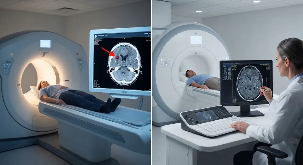
Quick answer: Neither scan is “better” in every case. A CT scan is usually the first choice in emergencies, such as a sudden severe headache, head injury or suspected stroke, because it takes only minutes and quickly shows bleeding and fractures.When considering which is better mri or ct scan for brain, an MRI is usually better for detailed brain evaluation, including small strokes, multiple sclerosis, tumors, seizures, inflammation and problems at the base of the brain. MRI uses no ionizing radiation but takes longer and is not safe for every implant.
Your doctor chooses based on what they are looking for, how urgent the situation is, and your medical history. This guide explains how that decision is made so you know what to expect.
The Short Version: CT vs MRI for the Brain
| Factor | Head CT | Brain MRI |
|---|---|---|
| Scan time | Usually under 5 minutes in the scanner | Often 30-60 minutes |
| Technology | X-rays and computer processing | Strong magnetic field and radio waves |
| Ionizing radiation | Yes | No |
| Best at showing | Fresh bleeding, skull fractures, large strokes, hydrocephalus | Brain tissue detail, small or early strokes, MS lesions, tumors, inflammation, seizure causes |
| Emergency use | First-line in most emergency rooms | Used when more detail is needed after initial assessment |
| Noise and enclosure | Quiet, open ring | Loud, longer tunnel (open and wide-bore designs exist) |
| Implants and metal | Rarely a problem | Some devices are unsafe or need special protocols |
| Contrast agent | Iodine-based (if needed) | Gadolinium-based (if needed) |
| Typical cost | Usually lower | Usually higher |
How a Head CT Works
A computed tomography (CT) scanner rotates an X-ray source around your head and builds cross-sectional images. Bone shows up bright white, and fresh blood usually appears brighter than surrounding brain tissue. That contrast is why CT is so good at answering urgent questions quickly:
Need to Schedule a Diagnostic Appointment?
Whether you need an MRI, ultrasound, X-ray, or another diagnostic service, Advantage MRI is here to help you take the next step in your healthcare journey.
Submit your appointment request online, and our team will contact you to confirm your visit.
Book Your Appointment →- Is there bleeding inside or around the brain?
- Is the skull fractured?
- Is there swelling or a shift of brain structures?
- Is there a large stroke or a blocked fluid pathway (hydrocephalus)?
A standard head CT uses a moderate dose of ionizing radiation. RadiologyInfo, a resource from the American College of Radiology and the Radiological Society of North America, places the effective dose of a head CT at about 2 millisieverts, which is comparable to several months of natural background radiation. For a single scan in a clear medical situation, the benefit generally outweighs the small risk. Doctors are more cautious about repeated scans, especially in children.
How a Brain MRI Works
Magnetic resonance imaging (MRI) uses a strong magnet and radio waves to create detailed images of soft tissue. Different MRI “sequences” highlight different features. For example:
- Diffusion-weighted imaging (DWI) can show an ischemic stroke very early, often before it is visible on CT.
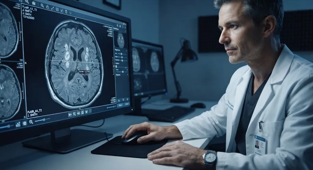
- FLAIR sequences highlight white matter lesions, such as those seen in multiple sclerosis.
- Gadolinium-enhanced sequences help show tumors, infection and active inflammation.
- Susceptibility-weighted imaging (SWI) can show tiny areas of past bleeding.
- MR angiography (MRA) can image blood vessels, often without contrast.
MRI does not use ionizing radiation, which makes it attractive for repeated follow-up scans, younger patients and pregnancy when imaging is truly needed. The trade-offs are time, noise, cost and safety screening for metal and implanted devices. If noise worries you, our article on why MRI scans are so loud explains what you’ll hear and how to cope.
When Is CT Better for the Brain?
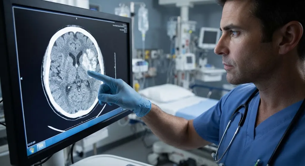
CT is usually the preferred first test when speed matters most.
Suspected Stroke in the First Hours
When someone arrives at an emergency department with stroke symptoms, a non-contrast head CT is typically done right away. Its main job is to rule out bleeding, because clot-busting medication can only be given if there is no hemorrhage. CT angiography may follow to look for a blocked large artery. MRI may be added later for more detail.
Head Injury
After trauma, CT quickly shows skull fractures and bleeding that might need surgery. Its speed also matters when a patient is unstable.
Sudden, Severe “Thunderclap” Headache
A sudden, worst-ever headache can signal bleeding around the brain (subarachnoid hemorrhage). CT is usually the first imaging test, often done in an emergency room.
Patients Who Can’t Have MRI
People with certain pacemakers, implanted devices or metal fragments, or patients who cannot lie still for a long time, may be imaged with CT instead.
Looking at Bone
CT shows bony detail of the skull, sinuses and inner ear very well.
Important: If you or someone near you has stroke symptoms, call 911 right away. Use the BE FAST signs: Balance loss, Eyes (vision change), Face drooping, Arm weakness, Speech difficulty, Time to call 911. Do not schedule outpatient imaging for a possible stroke.
When Is MRI Better for the Brain?
MRI is usually preferred when doctors need detailed information and the situation is not an immediate emergency.
Small or Early Strokes and TIAs
MRI with diffusion imaging is more sensitive than CT for small strokes, strokes in the brainstem and cerebellum, and strokes in the first hours. It can also help evaluate transient ischemic attacks (TIAs).
Multiple Sclerosis
MRI is central to diagnosing and monitoring MS. It shows lesions in the brain and spinal cord that CT usually cannot see.
Brain Tumors
MRI, especially with contrast, shows a tumor’s size, location and relationship to nearby structures. It is used for diagnosis, surgical planning and follow-up.
Seizures and Epilepsy
Specialized MRI protocols can find subtle structural causes of seizures, such as scarring or developmental abnormalities.
Chronic or Changing Headaches
Most headaches do not need imaging at all. When a doctor does order imaging for headaches with concerning features, such as new neurological symptoms or a change in pattern, MRI usually gives more information than CT.
Infection and Inflammation
Conditions such as encephalitis, abscesses and inflammatory diseases are better characterized with MRI.
The Back of the Brain (Posterior Fossa)
The brainstem and cerebellum sit near dense bone at the base of the skull. On CT, that bone creates streak artifacts that hide detail. MRI is not affected in the same way.
Dementia and Memory Problems
MRI can show patterns of brain shrinkage, small vessel disease and other changes that help a specialist evaluate memory loss. In some cases, a PET scan is added to look at brain activity. Our PET vs CT vs MRI guide explains how the three compare.
Brain MRI vs Head CT by Condition
| Condition or question | Usual first choice | Why |
|---|---|---|
| Head injury in the emergency room | CT | Fast; shows fractures and bleeding |
| Suspected acute stroke | CT first, MRI often follows | CT rules out bleeding quickly; MRI shows early or small strokes |
| Sudden worst-ever headache | CT | Detects bleeding around the brain |
| Multiple sclerosis | MRI | Shows white matter lesions CT misses |
| Brain tumor evaluation | MRI with contrast | Superior soft-tissue detail |
| New seizures | MRI | Finds subtle structural causes |
| Dizziness or brainstem symptoms (non-emergency) | MRI | Better view of the posterior fossa |
| Sinus disease | CT | Excellent bone and air detail |
| Memory loss evaluation | MRI | Shows atrophy and vascular changes |
These are typical patterns, not fixed rules. Your doctor may choose differently based on your situation.
Safety: Radiation, Contrast and Implants
Radiation
CT uses ionizing radiation. MRI does not. For a single medically justified head CT, the risk is considered small. If you have had many CT scans, mention it to your doctor so they can factor it into future decisions.
Contrast Agents
- CT contrast is iodine-based. It can cause allergic-type reactions in some people and may affect kidney function in people with significant kidney disease.
- MRI contrast is gadolinium-based. Serious reactions are uncommon. People with severe kidney disease need special consideration.
Many brain scans, especially emergency head CTs, are done without contrast.
Metal and Implanted Devices
MRI safety screening is essential. Tell the MRI team about:
- Pacemakers or defibrillators
- Cochlear implants
- Aneurysm clips
- Neurostimulators or implanted pumps
- Metal fragments, especially in the eyes
- Recent surgery with implanted hardware
Many modern devices are “MR conditional,” meaning they can be scanned safely under specific conditions. The imaging team needs the exact device information to decide.
Pregnancy
MRI without contrast is generally preferred over CT when brain imaging is necessary during pregnancy. Gadolinium is usually avoided unless clearly needed. Always tell the staff if you are or might be pregnant.
Claustrophobia
MRI takes longer inside a scanner. If you’re anxious in enclosed spaces, tell your doctor before the appointment. Options may include a mild sedative prescribed by your doctor, music, eye covers, or a facility with a wider-bore scanner.
What to Expect During a Brain MRI
- Screening. You’ll complete a safety questionnaire about metal, implants and medical history.
- Changing. You’ll remove jewelry, hairpins, hearing aids and anything metallic, and may change into a gown.
- Positioning. You’ll lie on your back with your head in a head coil, a cage-like device that helps capture clear images.
- Scanning. The scanner makes loud knocking and buzzing sounds. You’ll get earplugs or headphones. Staying still is the most important thing you can do for image quality.
- Contrast (if ordered). A small IV injection may be given partway through.
- After the scan. Most people return to normal activities right away unless they had sedation.
For eating, drinking and medication questions before your scan, see our guide on whether you can eat before an MRI.
Brain MRI With vs Without Contrast
Not every brain MRI needs contrast. The decision depends on the clinical question.
| Usually without contrast | Usually with contrast (or both) |
|---|---|
| Suspected stroke follow-up | Known or suspected brain tumor |
| Dementia and memory evaluation | Multiple sclerosis evaluation and some follow-ups |
| Many headache workups | Suspected infection or abscess |
| Hydrocephalus follow-up | Pituitary gland evaluation |
| Routine MRA of blood vessels in many cases | Inflammation of the brain lining (meningitis or other causes) |
When contrast is used, images are usually taken before and after the injection so the radiologist can compare. The IV contrast highlights areas where the normal blood-brain barrier is disrupted, which helps separate active disease from older scars. Tell the team if you’ve had a reaction to gadolinium before, have significant kidney disease, or are pregnant or breastfeeding.
Understanding Your Brain Imaging Report
Radiology reports use specialized language. A few common terms:
- “Unremarkable” or “within normal limits” means nothing abnormal was seen.
- “Nonspecific white matter hyperintensities” are small bright spots on MRI. They are common with aging, migraines, high blood pressure and other conditions, and are often not serious. Your doctor decides whether they need follow-up.
- “Incidental finding” is something unrelated to the reason for your scan, such as a small sinus cyst. Many incidental findings need no action.
- “Mass effect” or “midline shift” describes pressure on brain structures and is usually urgent.
- “Restricted diffusion” on MRI is often seen in a recent stroke, but it has other causes too.
- “Recommend clinical correlation” means the radiologist is asking your doctor to compare the images with your symptoms and exam.
Don’t try to interpret a report alone from a patient portal. Bring it to your follow-up visit and ask what each finding means for you.
What to Expect During a Head CT
A head CT is quick. You lie on a table that slides through a wide, short ring. The scan itself often takes less than a minute, and the whole visit can be done in well under half an hour if contrast is not needed. You’ll need to remove metal objects from your head and neck area.
Cost and Insurance Considerations
In general, a head CT costs less than a brain MRI. Prices vary widely by facility, region and insurance plan. Hospital-based imaging often costs more than imaging at an independent outpatient center. Many insurance plans require prior authorization for MRI, so your doctor’s office may need to submit paperwork before you can schedule. If you are paying out of pocket, ask for a written self-pay price before your appointment.
Our in-depth MRI vs CT scan guide covers costs, safety and uses across the whole body, not just the brain.
Can I Choose Which Scan I Get?
The ordering clinician decides which test is medically appropriate. You can and should ask questions, such as:
- Why are you choosing this scan over the other?
- Will I need contrast?
- What would the other scan show that this one won’t?
- If the first scan is normal, will I need the other one?
Doctors often follow evidence-based guidance, such as the American College of Radiology Appropriateness Criteria, to match the right test to each clinical question. Choosing the right first test can save time, money and unnecessary exposure. If you are curious about scanning without symptoms, our article on full-body MRI screening looks at the evidence.
Brain MRI at Advantage MRI in Chicago
Advantage MRI is an outpatient imaging center at 2008 N Pulaski Rd in Chicago. We provide MRI imaging, including brain MRI when ordered by your physician, along with X-ray, ultrasound and cardiac and nerve testing. We do not perform CT scans. If your doctor orders a head CT, especially for an emergency, that test is typically done at a hospital or emergency department.
Our website states that our MRI studies are interpreted by sub-specialized, board-certified radiologists and that the center is ACR accredited. You can read more about our team on our About page. We work with referring physicians and accept insured, out-of-network and self-pay patients.
Frequently Asked Questions About Which is Better MRI Or Ct Scan For Brain
Is MRI or CT better for detecting a brain tumor?
MRI, usually with contrast, is generally better for detecting and characterizing brain tumors. CT may detect larger tumors or be used when MRI isn’t possible.
Why do ERs do a CT instead of an MRI for head injuries?
CT is fast, widely available around the clock and very good at showing bleeding and fractures, which are the most urgent concerns after trauma.
Can a CT scan miss a stroke?
Yes. Early ischemic strokes and small strokes, especially in the brainstem, may not appear on CT in the first hours. MRI with diffusion imaging is more sensitive.
How long does a brain MRI take?
Most brain MRIs take about 30 to 60 minutes inside the scanner, depending on the protocol and whether contrast is used.
Is a brain MRI safe?
For most people, yes. MRI does not use ionizing radiation. The main safety concerns are metal objects and certain implanted devices, which is why careful screening is done before every scan.
Will I need both a CT and an MRI?
Sometimes. A CT is often done first in an emergency, and an MRI may follow to get more detail. In non-urgent situations, MRI alone is often enough.
Does a brain MRI show headaches?
Headaches themselves don’t appear on imaging. A brain MRI looks for structural causes, which are uncommon in typical tension or migraine headaches.
Conclusion
For brain imaging, CT and MRI do different jobs. CT is the fast, first-line tool for emergencies like trauma, bleeding and suspected stroke. MRI gives far more detail of brain tissue and is the better choice for small strokes, MS, tumors, seizures and many chronic or unexplained neurological symptoms. Your doctor weighs urgency, the question being asked, and your safety factors to decide which scan comes first.
Has your doctor ordered a brain MRI? Request an appointment with Advantage MRI or call (773) 227-9777. Hours: Monday-Friday 9:00 AM-5:00 PM, Saturday 9:00 AM-1:00 PM. Follow us on Facebook and Instagram for patient tips.