Health & Imaging
Echocardiogram vs EKG: What Sets These Heart Tests Apart
If your doctor just ordered an EKG and mentioned an echocardiogram in the same breath you are probably wondering if these are the same thing with two different names. They are not. And that mix up is more common than you would think.
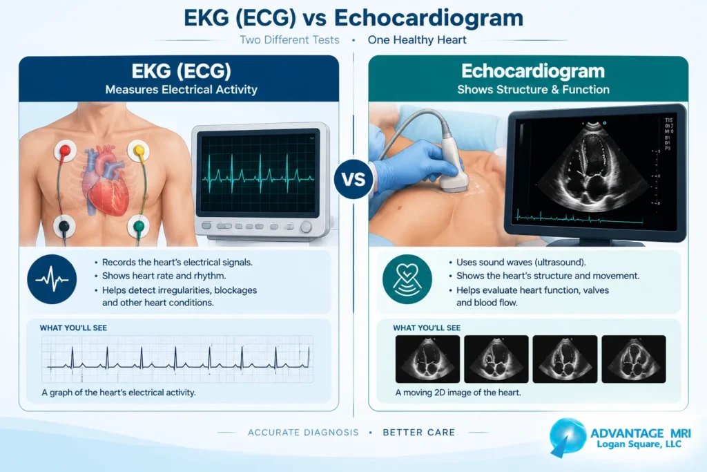
Here is the thing. Both tests look at your heart but they answer completely different questions. When comparing an echocardiogram vs ekg, one reads the electrical signals that make your heart beat. The other uses sound waves to show how your heart actually looks and moves. Once you understand that split the rest of it starts to make sense pretty fast.
This article breaks down what each test does how they differ what they can and cannot catch and when a cardiologist might order both. We will keep things simple and skip the jargon where we can.
What Is an EKG (ECG)
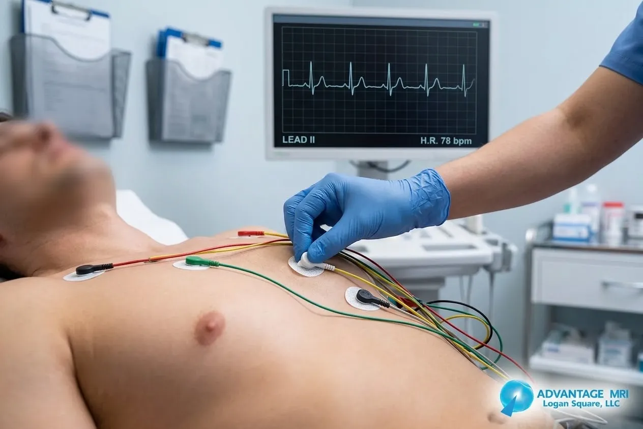
An EKG, also called an ECG or electrocardiogram, measures the electrical activity of your heart. Every heartbeat starts with a tiny electrical impulse that tells your heart muscle when to contract and when to relax. The EKG records that pattern using small electrode stickers placed on your chest arms and legs.
The test itself is quick and painless. You lie down, a technician attaches the electrodes and the machine prints out a wavy line graph showing your heart’s rhythm. Most EKGs take less than ten minutes start to finish.
How EKG Reads Electrical Activity
Each spike and dip on that graph represents a different part of your heartbeat. A cardiologist looks at the spacing, height and shape of those waves to figure out if your heart rhythm is regular or if something looks off. This is why an EKG is often the first test ordered when someone shows up with chest pain, palpitations or shortness of breath.
What EKG Can Detect
An EKG is good at picking up arrhythmia, which means an irregular heartbeat. It can also flag signs of a past heart attack, certain electrolyte imbalances and some structural heart disease if the electrical pattern is unusual enough. Some patients wear a continuous ECG device like a Holter monitor for a day or more if a single reading in the office does not catch anything unusual.
What it cannot do is show you a picture of your heart. It only reads electrical signals. So if your doctor needs to see the actual size shape and pumping strength of your heart, an EKG alone will not cut it.
What Is an Echocardiogram
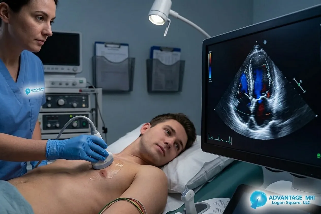
An echocardiogram uses ultrasound, the same sound wave technology used for imaging during pregnancy, to create detailed images of the heart. A sonographer moves a small wand called a transducer across your chest and the sound waves bounce off your heart structures to build a live moving picture on a screen.
This test gives your cardiologist a real time picture of your heart’s structure and function. That includes the four chambers, the heart valves, the muscle walls and how well blood is flowing through everything.
How Echo Uses Sound Waves
The transducer sends high frequency sound waves into your chest. Those waves bounce back differently depending on what they hit, whether that is heart muscle, valve tissue or blood moving through a chamber. The machine turns those echoes into an image, which is where the name echocardiogram comes from.
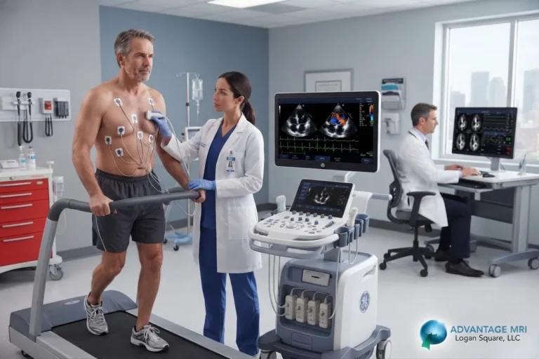
There are a few types worth knowing. A transthoracic echocardiogram is the standard version done from outside the chest. A transesophageal echocardiogram involves a small probe passed down the throat for a closer look at the heart, usually reserved for cases where a clearer image is needed. And a stress echocardiogram checks how your heart performs during physical exertion, often paired with a treadmill or medication that mimics exercise.
What Echo Shows About Heart’s Structure
An echo can measure how well your heart is pumping, a number often reported as ejection fraction. It can spot valve problems like leaking or narrowing, detect fluid around the heart and identify areas of heart muscle that are not moving normally, which sometimes points to reduced blood flow. It gives a picture of your heart’s health that an EKG simply cannot provide.
Echocardiogram vs EKG: Differences
| Feature | EKG (ECG) | Echocardiogram |
|---|---|---|
| What it measures | Heart’s electrical activity | Heart’s structure and function |
| Technology used | Electrodes and electrical signals | Ultrasound and sound waves |
| Typical duration | 5 to 10 minutes | 30 to 60 minutes |
| Shows heart rhythm | Yes | Limited |
| Shows valve function | No | Yes |
| Shows pumping strength | No | Yes |
| Invasive | No | No (transesophageal version is semi invasive) |
| Common use | Chest pain, palpitations, rhythm checks | Valve disease, heart failure, structural concerns |
EKG vs ECG Terminology Clarified
If you have seen both terms and wondered if EKG and ECG are different tests, they are not. EKG comes from the German spelling elektrokardiogramm and ECG is the English abbreviation. Doctors in the United States use both terms interchangeably to mean the exact same test.
What Each Test Can and Cannot Show
Can EKG Show Blockage
Not directly. An EKG can sometimes hint at reduced blood flow if the heart muscle’s electrical pattern is affected, especially during an active heart attack. But it cannot directly visualize a blocked artery. For that a cardiologist usually needs additional imaging or a cardiac stress test.
Can Echo Detect Heart Attack
An echocardiogram can show areas of heart muscle that are not contracting properly, which can be a sign of damage from a past or current heart attack. It is not typically the first test used to diagnose an active heart attack though. That job usually falls to the EKG combined with blood tests, with echo following up afterward to check for damage.
Is an Echocardiogram the Same as an EKG
No, they are not the same test. An echocardiogram uses ultrasound to create images of your heart’s structure, while an EKG uses electrodes to record your heart’s electrical activity. Think of it this way. The EKG tells you if your heart is beating in the right rhythm. The echocardiogram shows you what your heart actually looks like while it beats. Both matter but they answer different questions about your heart health.
When a Cardiologist Orders Both Tests Together
It is pretty common for a doctor to order an EKG and an echocardiogram at the same visit, especially if you come in with chest pain, shortness of breath or a history of heart disease. The EKG gives a quick snapshot of your heart’s rhythm right then and there. If anything looks unusual or if your doctor wants a fuller picture regardless, the echocardiogram follows up with a look at your heart’s structure.
For a lot of people this combination gives a much more complete picture than either test alone. An abnormal EKG paired with a normal echo tells a different story than an abnormal EKG with an abnormal echo and your cardiologist will use both pieces to decide next steps.
What to Expect During Each Test
EKG Test Prep and Duration
There is not much prep involved. You will be asked to lie still while small electrode patches are placed on your chest, arms and legs. The machine records for a minute or two and that is it. No needles, no discomfort, nothing invasive at all.
Echocardiogram Test Prep and Duration
An echocardiogram takes a bit longer, usually somewhere between 30 and 60 minutes depending on what your cardiologist is looking for. You will lie on your side while a sonographer moves the transducer across your chest with a bit of gel to help the sound waves transmit clearly. It is painless, though you might feel light pressure from the wand. No fasting or special prep is typically needed for a standard transthoracic echocardiogram.
Which Heart Test Do You Need
Signs You May Need an EKG
If you are dealing with chest pain, heart palpitations, dizziness or unexplained shortness of breath, your doctor will likely start with an EKG. It is fast, painless and gives an immediate look at your heart rhythm.
Signs You May Need an Echocardiogram
If your doctor suspects a valve problem, heart failure or wants a closer look at your heart’s pumping function, an echocardiogram is the better tool. It is also often used to monitor known heart conditions over time or check for changes after a cardiac event.
Your doctor will decide which test fits your situation and sometimes that means both. This article is not a substitute for medical advice and any decision about testing should come from a conversation with your physician.
Other Diagnostic Tests at Advantage MRI
Heart health is only part of the picture when it comes to diagnostic imaging. Advantage MRI in Chicago’s Hermosa neighborhood offers a full range of tests beyond ekg testing and echocardiogram services.
If your doctor is looking into nerve or muscle related symptoms, an emg testing or nerve conduction study session may be recommended. For ongoing heart rhythm monitoring over a longer stretch of time, a holter monitor can track your heart’s activity for a full day or more. And for broader imaging needs, our center also provides mri imaging, digital x ray and heart ultrasound imaging services, all read by our board certified radiologists.
FAQs
What is the difference between EKG and echocardiogram?
An EKG records your heart’s electrical activity using electrodes on your skin, while an echocardiogram uses ultrasound to create images of your heart’s structure and how it moves.
Which test is more accurate for heart problems?
Neither test is more accurate across the board. They measure different things. An EKG is better for catching rhythm issues, while an echocardiogram is better for structural problems like valve disease or weak pumping function.
Can EKG detect heart failure?
An EKG alone usually cannot confirm heart failure, though certain electrical patterns may raise suspicion. An echocardiogram is typically needed to measure pumping strength and confirm a heart failure diagnosis.
Does echocardiogram show blockages?
Not directly. An echocardiogram can show areas of heart muscle affected by reduced blood flow but it does not visualize the blocked artery itself the way other imaging tests might.
Is echocardiogram painful?
No. A standard transthoracic echocardiogram is painless. You may feel slight pressure from the ultrasound wand but there is no needle or incision involved.
How long does an EKG take?
A standard EKG takes about five to ten minutes including setup time for the electrodes.
Can I have a normal EKG but abnormal echo?
Yes, this happens fairly often. Your heart’s electrical rhythm can look normal on an EKG even if there is an underlying structural issue that only shows up on an echocardiogram, like a leaking valve or reduced pumping function.
Do I need both tests on the same day?
It depends on your symptoms and your doctor’s judgment. Many patients do have both tests done during the same visit, especially when chest pain or heart failure symptoms are involved.
Is an echocardiogram the same as an ultrasound of the heart?
Yes. An echocardiogram is essentially a heart specific ultrasound. It uses the same sound wave technology used in other ultrasound imaging, just focused on the heart’s chambers, valves and muscle walls.
How often should heart tests be repeated?
This varies based on your health history and any existing heart condition. Someone with a known valve problem or heart failure may need echocardiograms repeated periodically, while a person with no history of heart disease may only need testing if new symptoms come up. Your cardiologist can guide you on the right schedule.
Conclusion
An EKG and an echocardiogram are not interchangeable, even though they often get grouped together as heart tests. One reads the electrical signals that keep your heart beating in rhythm. The other creates a moving picture of your heart’s structure and function using sound waves. Doctors often lean on both together to get a fuller sense of your cardiovascular health, especially when symptoms like chest pain or shortness of breath show up. If your physician has ordered either test or both, that is a normal step toward understanding what is going on with your heart.
This article is meant for general information and is not a substitute for professional medical advice, diagnosis or treatment. Always talk to your doctor about your specific symptoms and testing needs.
For reliable, advanced diagnostic imaging you can trust, call Advantage MRI today or schedule your appointment online.
Contact Us
- Address: 2008 N Pulaski Rd, Chicago, IL 60639
- Phone: (773) 227 9777
- Contact us for more information
Follow us on social media for the latest health tips, diagnostic imaging updates and clinic news: Facebook | Instagram | LinkedIn.