
Lower back pain that won’t quit is frustrating. You rest it, you stretch it, you try to ignore it and it still there weeks later. At some point your doctor might mention an EMG test on lower back muscles and nerves to figure out what actually going on underneath the pain. If you’ve never had one before, the name alone sounds intimidating. It isn’t as complicated as it sounds and knowing what to expect makes the whole thing a lot less stressful.
This guide walks through what an EMG test on lower back nerves and muscles actually involves, why a doctor would order one, what happens step by step and what the results can tell you. We’ll also cover how it different from a nerve conduction study, since the two often get done together.
A lot of people searching for an emg test back option are already deep into weeks of pain, appointments and not much resolution. That normal. Nerve related back pain can take longer to diagnose than a simple muscle strain because the source of the pain isn’t always where the pain is felt. Someone can have a compressed nerve root in the lumbar spine and feel most of the pain running down their leg instead of in their back. Electromyography and nerve testing exist specifically to sort out that kind of confusion.
What Is an EMG Test
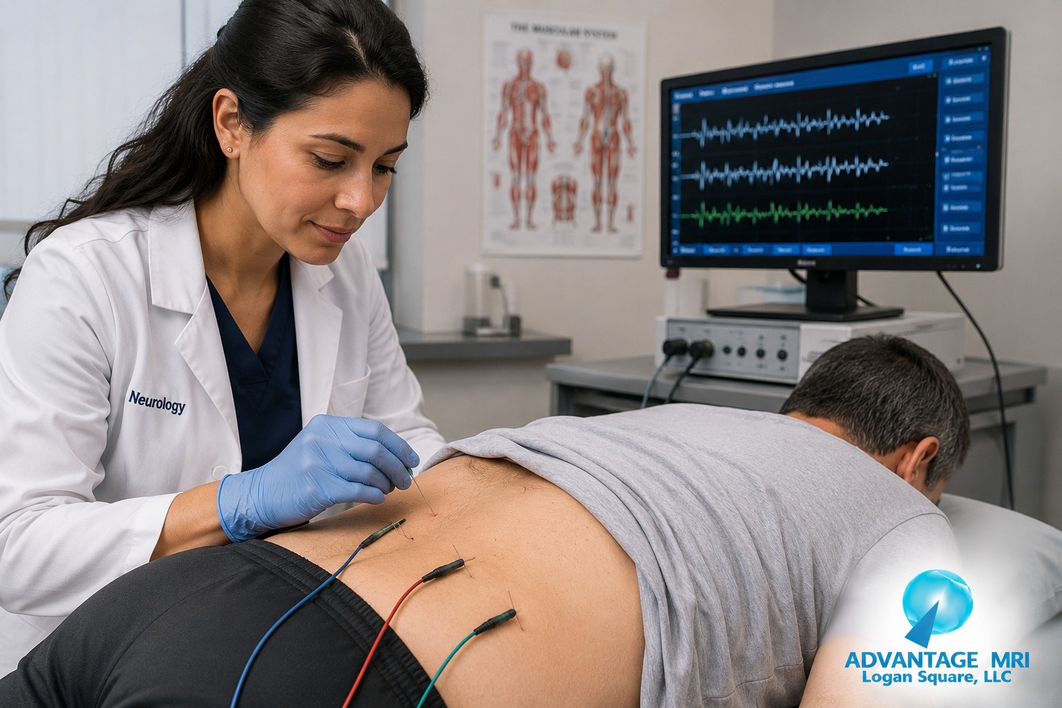
EMG stands for electromyography. In plain terms, it a test that checks how well your muscles respond to nerve signals. A thin needle electrode gets placed into the muscle being tested and it picks up the electrical activity happening inside that muscle both at rest and when you contract it.
Here the thing. Muscles don’t move on their own. A nerve tells them when to fire and the muscle responds. When there damage somewhere along that nerve pathway, whether it from a herniated disc pressing on a nerve root or something else going on in the lumbar spine, the muscle doesn’t get a clean signal. An EMG test on lower back muscles picks up exactly where that breakdown is happening.
How It Reads Muscle and Nerve Signals
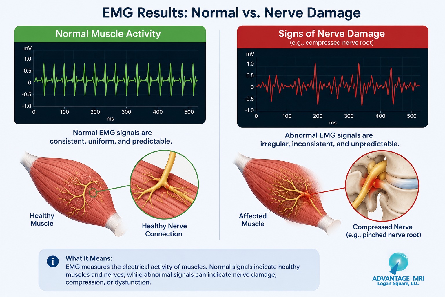
The needle electrode sends the electrical activity from your muscle to a monitor and a doctor trained in reading these signals looks for patterns. Normal muscle at rest is electrically quiet. Muscle that been affected by nerve compression often shows abnormal spontaneous activity even when you’re not trying to move it. That difference is what points toward nerve involvement instead of a simple muscle strain.
EMG is part of a broader category called electrodiagnostic studies and the peripheral nervous system is really what being examined here, not the brain and spinal cord directly. The peripheral nerves are the ones running from the spinal nerve root out through the lower back, hip and leg and they’re the ones most often affected in cases of low back pain with radiating symptoms. When someone mentions an emg test on legs or an emg test on feet, they’re usually describing the same electrodiagnostic process applied to a different part of the same nerve pathway, since a single lumbar nerve root can affect muscle groups all the way down to the foot.
Why Doctors Order EMG for Lower Back Pain
Not everyone with back pain needs this test. Most low back pain gets better on its own with rest, physical therapy and time. Doctors usually order an EMG test for back pain when there are signs that a nerve root itself might be involved rather than just sore muscles.
Common Symptoms That Lead to Testing
A doctor might recommend electromyography if you’re dealing with:
- Numbness or tingling that travels down the leg
- Muscle weakness in the leg or foot that doesn’t improve with rest
- Sciatica that hasn’t responded to conservative treatment
- Back pain paired with a change in reflexes
- Suspected nerve root compression from a herniated disc or spinal stenosis
If any of that sounds familiar, an EMG test combined with imaging can help your doctor build an actual treatment plan instead of guessing.
What Happens During the Test
The full process usually has two parts and most patients are surprised by how straightforward it is once they’ve been through it.
Before the test, you’ll talk with the technician or physician about your symptoms and medical history. There typically no need to fast or change your diet beforehand. You’ll be asked to wear loose clothing so the area being tested is easy to access.
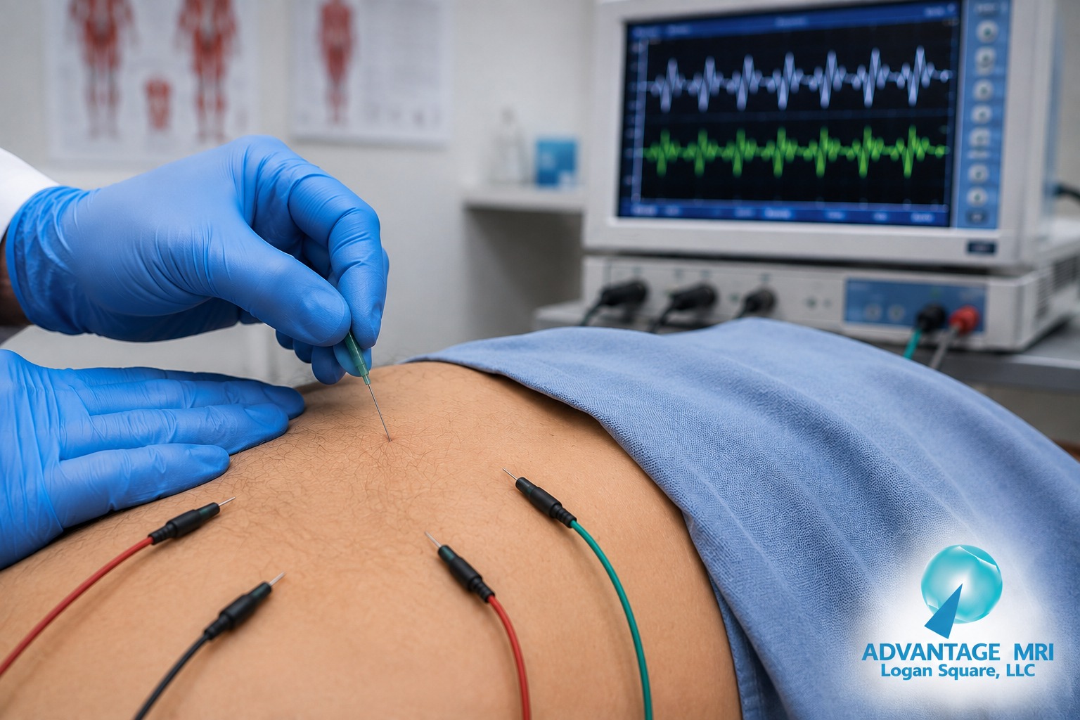
During the needle EMG portion, a thin needle electrode is placed into specific muscles in the lower back, hip or leg depending on which nerve root is suspected. You’ll be asked to relax the muscle and then gently contract it. The electrical readings at rest and during contraction get recorded and compared.
During the nerve conduction portion, small electrodes are placed on the skin instead of inserted and a mild electrical pulse is sent through the nerve to measure how fast and how strong the signal travels. This part measures nerve function directly rather than the muscle response to it.
The full appointment for an EMG test on lower back concerns typically takes between 30 and 60 minutes depending on how many muscle groups and nerves need to be checked. If symptoms extend down into the leg or foot, the technician may test additional muscle groups along that same nerve pathway, since a single lumbar nerve root often supplies more than one area. That part of why someone searching for an emg test on foot or an emg test on feet is often describing an extension of the same lower back workup rather than a completely separate test.
Anatomical location matters a lot here. The lumbar spine has several nerve roots and each one supplies a specific pattern of muscles down the leg. By testing the right combination of muscles, the physician reading the results can usually narrow down which exact nerve root is involved, sometimes down to a single spinal level.
EMG vs Nerve Conduction Study
These two tests get mentioned together so often that people assume they’re the same thing. They’re not, though they usually happen back to back as part of one appointment.
| EMG (Electromyography) | Nerve Conduction Study | |
|---|---|---|
| What it measures | Electrical activity inside the muscle | Speed and strength of nerve signals |
| How it done | Needle electrode inserted into muscle | Surface electrodes on skin |
| What it shows | Muscle response to nerve signals, nerve root damage | Nerve compression, peripheral neuropathy, carpal tunnel syndrome |
| Sensation | Brief pinch or ache during needle placement | Mild tingling or twitch from electrical pulse |
A doctor might order a nerve conduction study alongside the EMG when the goal is to see the full picture from nerve root all the way out to the peripheral nerve.
What Results Can Show
Results from an EMG test on lower back muscles fall into a few general categories and your doctor will walk you through what your specific findings mean for your situation.
Signs of Nerve Damage
Abnormal electrical activity at rest, delayed nerve signal timing or reduced signal strength can point toward nerve root compression, radiculopathy or peripheral nerve compression. This is often what confirms whether sciatica is coming from an actual pinched nerve versus muscle tightness.
Signs of Muscle Involvement
Sometimes the findings point more toward a primary muscle or neuromuscular condition rather than nerve compression. In rarer cases, EMG findings can be part of the workup for conditions like amyotrophic lateral sclerosis, though that a small fraction of why most people get tested. Muscle disease affecting the fibers directly looks different on EMG than nerve root damage does and that distinction alone can change the entire direction of a treatment plan. The same testing approach is also used outside the lower back for things like carpal tunnel syndrome, where nerve compression happens at the wrist instead of the spine, which shows how broadly electrodiagnostic testing applies across the body.
A normal EMG doesn’t always rule out a structural problem either. That part of why doctors often pair EMG findings with MRI imaging of the lumbar spine, since the MRI can show a herniated disc or spinal stenosis directly while the EMG shows how the nerve is actually functioning.
How to Prepare for Your EMG
There isn’t much prep involved, which is one less thing to worry about. A few things do help though:
- Avoid lotions or oils on the skin the day of testing
- Wear or bring loose clothing that allows easy access to the lower back and legs
- Let the technician know about any bleeding disorders or if you’re on blood thinners
- Mention any pacemaker or implanted device before the test starts
That really about it. There no fasting requirement and most people drive themselves home afterward.
Is an EMG Test Painful for Lower Back Testing
This is probably the question people ask most before their appointment. The needle electrode portion can feel like a brief pinch or a dull ache while it inserted and some people feel a mild cramping sensation when the muscle contracts. It not a pain free procedure but it also not something that lingers. Most patients describe it as uncomfortable rather than genuinely painful and any soreness afterward usually fades within a day.
The nerve conduction portion with surface electrodes causes a brief tingle or small muscle twitch from the electrical pulse. It over in a second and doesn’t leave any lasting discomfort.
Who Should Consider This Test
An EMG test on lower back nerves is generally recommended for people whose symptoms suggest actual nerve involvement rather than muscular strain. That includes people with radiating leg pain, unexplained weakness, numbness that follows a specific nerve pattern or back pain that hasn’t responded to typical treatment after several weeks.
It less commonly the first step for someone with simple, localized low back pain and no radiating symptoms. Your doctor will usually try conservative treatment or basic imaging first before recommending electrodiagnostic studies.
Chronic pain that sticks around for months tends to push doctors toward this kind of testing sooner rather than later. And that makes sense. The longer a nerve root stays compressed without a clear answer, the harder it can be to build an effective treatment plan. An EMG test on lower back or leg muscles gives a functional answer that a physical exam alone often can’t provide, especially when symptoms overlap with something like peripheral neuropathy, which can also cause tingling and weakness in the legs but comes from a different underlying cause than a compressed nerve root.
EMG Testing for Sciatica and Herniated Disc
Sciatica happens when the sciatic nerve gets irritated or compressed and a herniated disc in the lumbar spine is one of the most common causes. An EMG test can confirm whether the nerve root feeding the sciatic nerve is actually affected and it can help pinpoint which level of the spine the problem is coming from.
This matters for treatment planning. Knowing the exact nerve root involved helps guide decisions around physical therapy, injections or, in more involved cases, surgical consultation. Pairing the EMG with an MRI gives both the structural picture and the functional picture, which is often more useful together than either test alone.
What Happens After the Test
There no real recovery period. You might have mild soreness at the needle sites for a day, similar to a light bruise. Most people go back to normal activity right away. Results usually get reviewed by a physician trained in reading electrodiagnostic studies and findings get sent to the referring doctor to guide next steps in your treatment plan.
Frequently Asked Questions
Does an EMG test show lower back nerve damage?
Yes. It measures electrical activity in the muscle and can pick up abnormal patterns that point to nerve root compression or damage affecting the lower back and legs.
Is an EMG test painful for lower back testing?
It can cause brief discomfort from the needle electrode, similar to a pinch, along with a mild ache during muscle contraction. Most people tolerate it well and any soreness fades quickly.
How long do EMG results take?
This depends on the facility and the complexity of the findings but a physician typically reviews and interprets results within a short turnaround so your doctor can move forward with a treatment plan.
Can an EMG test detect sciatica?
It can help confirm whether sciatica symptoms are coming from actual nerve root compression rather than muscle tightness and it can help identify which nerve level is involved.
What is the difference between EMG and a nerve conduction study?
EMG measures electrical activity inside the muscle using a needle electrode. A nerve conduction study measures how fast and how strong a signal travels along the nerve using surface electrodes. They’re often done together.
Why would a doctor order an EMG for back pain?
Doctors typically order this test when symptoms suggest nerve involvement, such as radiating leg pain, numbness or muscle weakness that hasn’t improved with conservative treatment.
How do I prepare for an EMG test on lower back concerns?
Avoid lotions on the skin, wear loose clothing and let the technician know about blood thinners or implanted devices. No fasting is needed.
Is an EMG test covered by insurance?
Coverage depends on your specific insurance plan and whether the test is considered medically necessary based on your symptoms. Check with your provider or our office to confirm your coverage.
Can an EMG test diagnose a herniated disc?
An EMG shows how the nerve is functioning rather than the physical structure of the disc itself. A herniated disc is usually confirmed with imaging like an MRI, while the EMG shows whether that disc is actually affecting nerve function.
How accurate is an EMG for back pain?
When performed and interpreted by a qualified physician, EMG is a reliable tool for identifying nerve root involvement, though it works best alongside a full clinical picture and imaging rather than as a standalone answer.
Final Thoughts
An EMG test on lower back nerves and muscles gives your doctor real information about whether your pain is coming from nerve compression, a muscle issue or something else entirely. It not the first test for every case of back pain but for symptoms like radiating leg pain, numbness or unexplained weakness, it can be the piece that finally connects the dots. This information is meant to help you understand what to expect and isn’t a substitute for advice from your own physician.
At Advantage MRI in the Hermosa neighborhood of Chicago, testing is overseen with board certified expertise and read with the same level of care as our other diagnostic services, including ultrasound, echocardiogram, EKG testing, holter monitor and digital x ray. Our founder, Dr. Gregory T. Goldstein, is a board certified diagnostic radiologist with a background in orthopedic and neurologic imaging and that focus shapes how testing is handled here from start to finish. You can learn more about our practice or get in touch through our contact page.
For reliable, advanced diagnostic imaging you can trust, call Advantage MRI today or schedule your appointment online.
Contact Us
- Address: 2008 N Pulaski Rd, Chicago, IL 60639
- Phone: (773) 227 9777
Follow us on social media for the latest health tips, diagnostic imaging updates and clinic news: Facebook | Instagram | LinkedIn.