Health & Imaging
MRI vs X-Ray: Which Imaging Test Does Your Injury Actually Need?
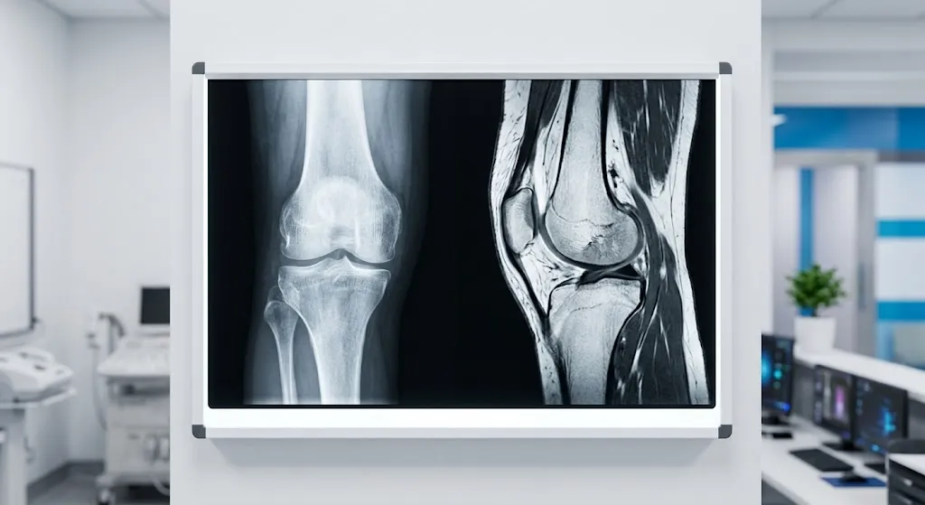
When comparing mri vs x ray, a quick answer is that an X-ray is a fast, low-cost test that’s best for looking at bones: fractures, dislocations, arthritis and alignment. It takes minutes and uses a small dose of radiation. An MRI uses magnets and radio waves, with no radiation, to show soft tissues in detail: ligaments, tendons, cartilage, muscles, spinal discs, nerves and bone marrow. For most injuries, doctors start with an X-ray and order an MRI only if the X-ray is normal but symptoms suggest a soft-tissue injury, or if more detail is needed for treatment.
This guide explains what each test shows, how doctors decide, why insurance often requires an X-ray first, and what to expect on test day. If you need imaging in Chicago, we also explain how to get both tests at one location.
MRI vs X-Ray at a Glance
| X-ray | MRI | |
|---|---|---|
| Technology | Small dose of ionizing radiation | Magnetic field and radio waves |
| Radiation | Yes (low dose) | None |
| Best for | Bones, fractures, joint alignment, arthritis, chest and lungs | Soft tissue: ligaments, tendons, cartilage, menisci, discs, nerves, muscles, bone marrow |
| Time | A few minutes | Often 20-60 minutes |
| Cost | Low | Higher |
| Availability | Often same-day or walk-in | Usually scheduled; may need prior authorization |
| Comfort | Quick, quiet, open | Loud, longer, inside a scanner |
| Metal implants | Not a safety issue | Must be screened |
| Typical role | First test for most injuries | Follow-up test for detail |
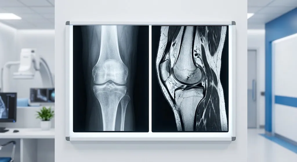
What an X-Ray Shows
An X-ray (radiograph) passes a small amount of radiation through your body onto a detector. Dense structures like bone block more X-rays and appear white. Air appears black. Soft tissues appear in shades of gray but with little detail.
Need to Schedule a Diagnostic Appointment?
Whether you need an MRI, ultrasound, X-ray, or another diagnostic service, Advantage MRI is here to help you take the next step in your healthcare journey.
Submit your appointment request online, and our team will contact you to confirm your visit.
Book Your Appointment →X-rays are excellent for:
- Fractures (broken bones)
- Dislocations and joint alignment
- Arthritis, shown by joint space narrowing and bone spurs
- Bone deformities and growth problems
- Some bone tumors and infections
- Foreign objects, such as metal fragments
- Chest conditions, such as pneumonia, fluid around the lungs and heart size
- Spinal alignment, including scoliosis and slippage of vertebrae
X-rays have limits. They don’t show ligaments, tendons, cartilage, menisci, discs or nerves directly. Some fractures, especially early stress fractures or hairline fractures in certain bones such as the scaphoid in the wrist, may not appear on the first X-ray.
Is X-Ray Radiation Safe?
The dose from most X-rays is low. A chest X-ray, for example, is commonly estimated at about 0.1 millisievert, roughly equal to around 10 days of natural background radiation. Extremity X-rays, like a hand or ankle, are usually even lower. Tell the technologist if you are or might be pregnant so they can decide whether to proceed and how to shield appropriately.
What an MRI Shows
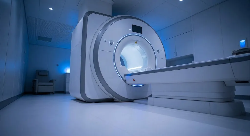
MRI creates detailed images using a strong magnet and radio waves. It excels at distinguishing between soft tissues, which is exactly where X-ray falls short. MRI is the preferred test for:
- Ligament tears, such as the ACL in the knee or ankle ligaments
- Meniscus tears in the knee
- Rotator cuff and labrum tears in the shoulder
- Tendon injuries, such as Achilles or hamstring tears
- Cartilage damage
- Herniated discs and nerve compression in the spine
- Spinal cord problems
- Stress fractures and bone bruises not visible on X-ray
- Muscle tears
- Soft-tissue masses
- Infection or inflammation in bones and joints
If MRI noise or the enclosed space concerns you, see our explanation of MRI scanning sounds and how to cope.
Why Do Doctors Usually Order an X-Ray First?
You may wonder why you need an X-ray if an MRI shows more. There are good reasons:
- It answers the most urgent question fast. Is a bone broken or dislocated? That changes treatment immediately.
- It’s quick, available and inexpensive.
- It can make MRI unnecessary. If the X-ray shows a clear fracture or advanced arthritis, an MRI may add little.
- It helps interpret the MRI. Radiologists often compare the two. For example, an X-ray shows arthritis severity and alignment while bearing weight, which MRI (done lying down) can’t show the same way.
- Guidelines recommend it. The American College of Radiology Appropriateness Criteria commonly list X-rays as the first imaging test for many joint and bone complaints.
- Insurance often requires it. Many insurers won’t approve an MRI for joint pain without a recent X-ray and a period of conservative treatment.
MRI or X-Ray: Which Test for Your Injury?
| Injury or symptom | Usual first test | When MRI may follow |
|---|---|---|
| Possible broken bone after a fall | X-ray | If X-ray is negative but pain or tenderness persists, or to assess surrounding soft tissue |
| Ankle sprain | X-ray if specific warning signs are present | If pain or instability lasts weeks, to check ligaments or hidden fractures |
| Knee “pop” with swelling | X-ray | To check for ACL, meniscus or cartilage injury |
| Shoulder pain with weakness | X-ray | To check rotator cuff or labrum tears |
| Wrist pain after falling on an outstretched hand | X-ray | If scaphoid fracture is suspected despite a normal X-ray |
| Low back pain without red flags | Often no imaging in the first weeks | If pain persists with leg symptoms or red flags develop |
| Back pain with leg numbness or weakness | MRI may be first, depending on findings | Shows discs and nerves |
| Neck pain after trauma | X-ray or CT, depending on severity | If nerve symptoms or ligament injury suspected |
| Runner with shin or foot pain | X-ray | To detect early stress fractures |
| Joint pain in older adults | X-ray | If X-ray doesn’t explain symptoms or before certain treatments |
| Hip pain | X-ray | For labral tears, stress fractures or early bone problems |
These are common patterns. Your doctor considers your history, exam findings and risk factors.
A Note on Back Pain
Most new low back pain improves within a few weeks without imaging. Guidelines generally advise against routine imaging in the first several weeks unless there are “red flags,” such as significant trauma, fever, unexplained weight loss, a history of cancer, progressive weakness, or loss of bladder or bowel control. Loss of bladder or bowel control or numbness in the groin area is an emergency. If back pain comes with leg numbness, tingling or weakness, nerve testing may also be considered. Our article on EMG testing for lower back, leg and foot pain explains how EMG complements MRI.
Can an X-Ray Miss Something an MRI Would Find?
Yes. That’s expected, not a failure. X-rays are designed for bones. A normal X-ray can still accompany:
- A torn ligament or tendon
- A meniscus or cartilage tear
- A herniated disc pinching a nerve
- An early stress fracture
- A bone bruise
- Inflammation of the bone marrow
If your symptoms don’t match a normal X-ray, tell your doctor. That’s often when an MRI becomes appropriate.
Can an MRI Show Fractures?
Yes. MRI can detect fractures, including subtle ones X-rays miss, because it shows the swelling inside the bone. However, X-ray or CT often shows fracture alignment and fine bone detail more clearly. For complex fractures, especially for surgical planning, CT may be used. Our MRI vs CT scan guide explains when CT fits in.
Common Scenarios, Explained
The table above is a quick reference. These examples show how the decision plays out in real life.
A twisted knee playing basketball. You heard a pop, the knee swelled within hours and it feels unstable. Your doctor examines the knee and orders X-rays to check for fractures, including small bone fragments that a torn ligament can pull away. The X-rays are normal. Because the history and exam suggest an ACL or meniscus tear, an MRI is ordered next. The MRI shows the ligament and meniscus directly and helps the orthopedic surgeon plan treatment.
Shoulder pain lifting overhead. Months of aching and weakness raising your arm lead to X-rays first. They may show bone spurs or arthritis that narrow the space for the rotator cuff tendons. If pain continues after physical therapy, or if the doctor suspects a full-thickness tear, an MRI shows the tendons themselves.
A fall on an outstretched hand. Wrist X-rays are taken. The scaphoid, a small bone near the thumb side of the wrist, can break without showing up on the first X-ray. If you’re tender in that spot, your doctor may immobilize the wrist and either repeat X-rays later or order an MRI to detect the fracture early. Missing this fracture matters because the bone has a fragile blood supply.
A runner with foot pain. Pain started gradually after increasing mileage. Early stress fractures often don’t appear on X-ray for a couple of weeks. MRI can show bone stress changes much earlier, which helps guide how long to rest.
Hip pain in an older adult. X-rays are usually enough to show arthritis and guide treatment. MRI is reserved for cases where X-rays don’t explain the pain or a hidden fracture is suspected after a fall.
Why Weight-Bearing X-Rays Still Matter
MRI is done lying down, and most MRI scanners can’t show how a joint behaves under your body weight. Standing X-rays of the knees, feet, ankles or spine show joint space narrowing, alignment and deformities as they really are when you’re upright. That information guides decisions such as bracing, injections or joint replacement. It’s one more reason an X-ray is often ordered even when an MRI is planned.
Metal Implants: X-Ray vs MRI
If you have a joint replacement, plates, screws or rods from a previous surgery, X-ray is often the main follow-up tool. It shows implant position, loosening and healing well, and metal poses no safety issue.
MRI can still be done in many people with orthopedic hardware. Most modern orthopedic implants are considered safe in MRI, but the metal can create image distortion near the implant. Many centers use special metal-reduction sequences to improve image quality. Certain other devices, such as some pacemakers, neurostimulators and older aneurysm clips, need specific review before any MRI. Always bring your implant card or surgical records so the MRI team can check your device and decide whether and how to scan.
What to Expect During an X-Ray
- You’ll remove jewelry or clothing with metal near the area being imaged and may wear a gown.
- The technologist positions the body part on or in front of the detector. You may be asked to stand, sit or lie down, and to hold very still or hold your breath briefly.
- Usually two or more views are taken from different angles.
- The whole process typically takes 5 to 15 minutes.
There’s no special preparation for most X-rays. You can return to normal activities right away.
What to Expect During an MRI
- You’ll complete a safety screening questionnaire about implants, metal and medical history.
- You’ll remove metal items and may change into a gown.
- You’ll lie on a padded table. A coil may be placed around the body part being scanned.
- The table slides into the scanner. You’ll hear loud knocking sounds and receive earplugs or headphones.
- Each sequence lasts a few minutes; the entire exam often takes 20 to 60 minutes.
- Some exams use an IV contrast injection.
Food and drink rules depend on the exam. See our guide on eating before an MRI.
Cost: MRI vs X-Ray
An X-ray is one of the least expensive imaging tests. An MRI costs considerably more because of equipment, time and interpretation complexity. Costs vary by facility type, insurance plan and whether contrast is used. Independent outpatient centers often charge less than hospital outpatient departments. Ask for an estimate and check whether prior authorization is needed before scheduling an MRI.
MRI vs X-Ray vs CT vs PET
X-ray and MRI are two of several imaging tools. CT provides fast cross-sectional images with strong bone and lung detail, and PET shows how tissues function. For an overview of the full picture, read our PET vs CT vs MRI guide.
Getting an X-Ray in Chicago
If you need an X-ray in Chicago, look for:
- A facility that accepts your insurance or offers clear self-pay pricing
- Convenient hours and short wait times
- Board-certified radiologist interpretation
- Digital X-ray, which produces images quickly and lets results be shared electronically with your doctor
- On-site MRI, so follow-up imaging, if needed, can be done at the same place
Advantage MRI provides digital X-ray and MRI imaging at 2008 N Pulaski Rd, Chicago, IL 60639, serving the Hermosa, Logan Square and Humboldt Park neighborhoods and patients across Chicago. Having both tests under one roof means your doctor can order an X-ray first and, if needed, an MRI at the same facility, with images and reports available together. Most X-rays require a doctor’s order. Call ahead if you’re unsure what you need.
We welcome insured, out-of-network and self-pay patients.
Frequently Asked Questions
What is the main difference between MRI and X-ray?
X-rays use a small dose of radiation and are best for bones. MRI uses magnets and radio waves, has no radiation, and shows soft tissues such as ligaments, tendons, cartilage, discs and nerves.
Why did my doctor order an X-ray instead of an MRI?
X-rays are fast, inexpensive and answer the first key question: is there a fracture, dislocation or arthritis? Guidelines and insurers often require an X-ray before MRI.
Can an X-ray show a torn ligament?
Not directly. An X-ray may show indirect signs, such as a small bone fragment pulled off by a ligament or joint misalignment. MRI is needed to see the ligament itself.
Is MRI better than X-ray for back pain?
For back pain with nerve symptoms, MRI is more useful because it shows discs and nerves. For uncomplicated back pain, often no imaging is needed early on.
Which is safer, MRI or X-ray?
Both are safe for most people. X-rays use a low dose of radiation. MRI uses no radiation but requires screening for metal and implants.
How long does an X-ray take compared to an MRI?
An X-ray usually takes 5 to 15 minutes. An MRI typically takes 20 to 60 minutes in the scanner.
Can I get an X-ray in Chicago without an appointment?
Policies vary. Most facilities require a doctor’s order. Call Advantage MRI at (773) 227-9777 to check availability and requirements.
Conclusion
X-ray and MRI aren’t competitors. They work in sequence. X-ray is the fast, low-cost first look at bones and joints, and it often answers the question. MRI follows when soft tissue needs a closer look or when symptoms don’t match a normal X-ray. Knowing what each test shows helps you understand your doctor’s plan and what to expect.
Need an X-ray or MRI in Chicago? Book an appointment at Advantage MRI or call (773) 227-9777. Hours: Monday-Friday 9:00 AM-5:00 PM, Saturday 9:00 AM-1:00 PM. Questions? Contact us. Follow us on Facebook and Instagram.