Health & Imaging
What Does an EMG Test Show? Conditions It Diagnoses and How to Read Results
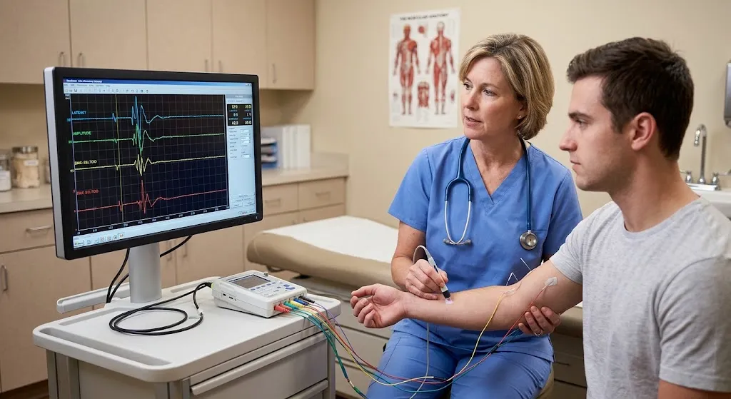
Quick answer: An EMG (electromyography) test shows how well your muscles and the nerves that control them are working. By recording the electrical activity of muscles at rest and during contraction, it helps doctors tell whether weakness, numbness, tingling or pain comes from a nerve problem (such as a pinched nerve in the neck or back, carpal tunnel syndrome or peripheral neuropathy), a muscle disease (myopathy), or a problem where nerve and muscle meet. An EMG is usually done together with a nerve conduction study (NCS), which measures how fast and how strongly signals travel along nerves. Together they can show where a problem is, how severe it is, and whether it’s new or longstanding.
This guide explains what an EMG measures, which conditions it helps diagnose, what “positive” or abnormal results mean, and what it can’t tell you.
Need to Schedule a Diagnostic Appointment?
Whether you need an MRI, ultrasound, X-ray, or another diagnostic service, Advantage MRI is here to help you take the next step in your healthcare journey.
Submit your appointment request online, and our team will contact you to confirm your visit.
Book Your Appointment →What Is an EMG Test?
Electrodiagnostic testing usually has two parts:
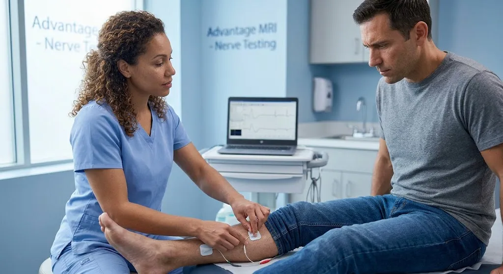
- Nerve conduction study (NCS): Small sticky electrodes are placed on the skin. Brief, mild electrical pulses stimulate a nerve, and the response is recorded. This measures the speed and strength of nerve signals.
- Needle EMG: A very thin needle electrode is inserted into selected muscles to record their electrical activity at rest and when you gently tighten them.
The term “EMG test” is often used for the whole visit. For a clear breakdown of the two parts, read EMG vs nerve conduction study.
What Does an EMG Test Show? The Core Questions It Answers
An EMG helps answer specific questions that imaging alone often can’t:
- Is the problem in the nerve or the muscle?
- Where along the nerve is the problem? For example, at the wrist, elbow, neck or lower back.
- Is it damage to the nerve’s insulation (myelin) or to the nerve fiber itself (axon)?
- How severe is it?
- Is it new (acute) or old (chronic)? And is there evidence of recovery?
- Is it affecting one nerve, several nerves, or nerves throughout the body?
MRI shows anatomy, such as a disc bulging toward a nerve. EMG shows function: whether that nerve is actually being affected. That’s why the two tests are often used together.
What Is an EMG Test Used to Diagnose?
Pinched Nerves in the Neck or Back (Radiculopathy)
A nerve root can be compressed where it leaves the spine, often by a herniated disc or narrowing of the spinal canal. This can cause pain, numbness or weakness radiating down an arm or leg. EMG can show which nerve root is involved and how much it’s affected. Learn more in our guide to EMG testing for lower back, leg and foot pain.
Carpal Tunnel Syndrome
Compression of the median nerve at the wrist is one of the most common reasons for testing. Nerve conduction studies typically show slowed signals across the wrist, and EMG can show whether the thumb muscles have been affected. See EMG testing for carpal tunnel syndrome.
Other Entrapment Neuropathies
- Ulnar neuropathy at the elbow (cubital tunnel), causing numbness in the ring and little fingers
- Peroneal (fibular) neuropathy at the knee, which can cause foot drop
- Tarsal tunnel syndrome at the ankle
Peripheral Neuropathy
Damage to many nerves, often starting in the feet, is common with diabetes and can also relate to vitamin deficiencies, alcohol, certain medications, kidney disease and inherited conditions. Testing can show the pattern and whether the main problem is in the axons or the myelin.
Brachial Plexus and Lumbosacral Plexus Injuries
The nerve networks in the shoulder and pelvis can be injured by trauma, inflammation or other causes. EMG helps localize and grade these injuries.
Muscle Diseases (Myopathies)
Inflammatory muscle conditions, muscular dystrophies and other myopathies produce a characteristic EMG pattern that differs from nerve damage.
Neuromuscular Junction Disorders
Conditions such as myasthenia gravis affect the point where nerve signals pass to muscle. Specialized techniques, like repetitive nerve stimulation, are used when these are suspected.
Motor Neuron Disease
In conditions such as amyotrophic lateral sclerosis (ALS), EMG findings across several body regions are an important part of the diagnostic workup, which is always done by specialists alongside other evaluations.
Nerve Injuries From Trauma
After a cut, crush or stretch injury, EMG can show whether a nerve is partly or completely injured and later whether it’s recovering.
How EMG Results Are Read
Results are interpreted by a physician trained in electrodiagnostic medicine, often a neurologist or physical medicine and rehabilitation physician. The final report combines nerve conduction numbers and needle EMG observations.
What the Nerve Conduction Study Measures
| Measurement | What it means | What abnormal may suggest |
|---|---|---|
| Latency | Time for the signal to travel and produce a response | Slowing at a specific spot, such as carpal tunnel |
| Amplitude | Size of the response | Fewer working nerve fibers (axonal loss) |
| Conduction velocity | Speed of the signal along a nerve segment | Myelin damage (demyelination) |
| F-waves and H-reflexes | Responses that travel toward the spine and back | Problems closer to the spinal cord or nerve roots |
What the Needle EMG Looks For
| Finding | What it generally indicates |
|---|---|
| Normal resting silence | A healthy muscle is electrically quiet at rest |
| Fibrillation potentials and positive sharp waves | Spontaneous activity often seen when muscle fibers have lost nerve supply recently or are actively damaged |
| Fasciculations | Spontaneous twitches of a motor unit; can be benign or seen in certain nerve conditions |
| Large, long motor unit potentials with reduced recruitment | A “neurogenic” pattern, consistent with longstanding nerve damage and reinnervation |
| Small, short, polyphasic potentials with early recruitment | A “myopathic” pattern, consistent with muscle disease |
| Myotonic discharges | Characteristic of certain muscle disorders |
These are simplified descriptions. The physician interprets the overall pattern in the context of your symptoms and exam.
What Does a “Positive” EMG Test Result Mean?
People often search for “positive EMG test results.” In practice, reports rarely use the word “positive.” Instead, a report states whether the study is normal or abnormal and gives an interpretation, such as:
- “Electrodiagnostic evidence of moderate median neuropathy at the wrist, consistent with carpal tunnel syndrome.”
- “Findings consistent with a chronic left L5 radiculopathy.”
- “Evidence of a length-dependent sensorimotor polyneuropathy, primarily axonal.”
- “No electrodiagnostic evidence of neuropathy, radiculopathy or myopathy.”
An abnormal (sometimes called positive) result means the test found evidence of a nerve or muscle problem. The report usually describes:
- The type of problem: nerve, nerve root, plexus, muscle or junction
- The location
- The severity: mild, moderate or severe
- The timing: acute, chronic or both
- Signs of recovery, when relevant
Your referring doctor uses this information, along with imaging and your exam, to plan treatment.
What Does a Normal EMG Mean?
A normal EMG means no evidence of significant nerve or muscle dysfunction was found in the nerves and muscles tested. It can be reassuring, but it doesn’t rule out every problem.
What an EMG Can’t Show: Important Limitations
- Very early nerve injury. Some EMG changes take time to appear. After a nerve injury, spontaneous activity in muscles often isn’t visible for roughly two to three weeks. Testing too early can underestimate the problem. Your doctor may schedule testing with this timing in mind.
- Small-fiber neuropathy. Standard NCS and EMG mainly test large nerve fibers. Small-fiber neuropathy, which often causes burning pain, may need other tests, such as a skin biopsy.
- Purely sensory nerve root problems. A pinched nerve root that affects only sensation may not show up on EMG.
- Pain alone. EMG measures nerve and muscle function, not pain itself.
- Brain and spinal cord detail. EMG doesn’t image these structures; MRI does.
- Only the areas tested. The physician chooses which nerves and muscles to test based on your symptoms.
Factors That Can Affect EMG Results
- Cold limbs can slow nerve conduction, which is why technicians may warm your hands or feet.
- Age and height affect normal values.
- Swelling can make recordings harder.
- Lotions can interfere with electrode contact.
- Ability to relax and contract muscles on request.
Simple steps before the test help. See our guide on EMG test preparation.
Symptoms That Commonly Lead to an EMG
Doctors usually order EMG and nerve conduction testing when symptoms point toward a nerve or muscle problem, such as:
- Numbness or tingling in the hands, fingers, feet or toes
- Pain that shoots or radiates down an arm or leg
- Weakness, such as dropping objects, trouble gripping or a foot that drags
- Muscle cramps, twitching or wasting (visible loss of muscle bulk)
- Unexplained muscle pain or fatigue with weakness
- Symptoms after an injury to an arm, leg, neck or back
These symptoms have many possible causes, including some that aren’t nerve-related. The EMG helps narrow them down.
Questions to Ask Your Doctor About Your EMG Results
- Was my study normal or abnormal?
- If abnormal, which nerve or muscle is affected, and where?
- How severe is it, and is it new or longstanding?
- Does it match my MRI or other test results?
- What treatment options fit these findings?
- Will I need a repeat EMG, and when?
How EMG Results Guide Treatment
An EMG report is more than a yes-or-no answer. Details about severity and timing often shape what happens next. A few common examples:
Carpal tunnel syndrome. Mild findings often lead to conservative care first, such as night splints, activity changes or, in some cases, a steroid injection. Severe findings, especially signs of nerve fiber loss in the thumb muscles, may prompt earlier discussion of surgical release with a hand specialist, because long-standing compression can lead to permanent weakness.
Pinched nerve in the lower back. If EMG shows an active, ongoing nerve root problem that matches the MRI and your symptoms, your doctor may consider physical therapy, medications, injections or a surgical opinion depending on severity. If EMG shows only old, stable changes, the current symptoms may have another explanation.
Peripheral neuropathy. The pattern of findings, such as axonal versus demyelinating, helps your doctor decide which blood tests or other evaluations to order to look for a cause.
Nerve injury after trauma. A follow-up EMG weeks to months later can show whether the nerve is regrowing, which helps surgeons decide whether to wait or operate.
When Should an EMG Be Done?
Timing matters. After a new nerve injury or a sudden pinched nerve, some EMG changes take roughly two to three weeks to appear. For that reason, doctors often wait a few weeks after symptoms start before ordering EMG, unless there’s a reason to test sooner, such as a baseline before surgery. For slowly developing conditions like carpal tunnel syndrome or neuropathy, testing can be done whenever symptoms are persistent enough to need evaluation.
Can an EMG Be Repeated?
Yes. Repeat studies are sometimes used to track recovery after nerve injury or surgery, to monitor progression of a neuropathy, or to re-evaluate when symptoms change. Bringing your previous EMG report helps the physician compare results.
EMG vs MRI: Which Does What?
| EMG/NCS | MRI | |
|---|---|---|
| Shows | How nerves and muscles function | Anatomy and structure |
| Example | Whether the L5 nerve is actually damaged | Whether a disc is touching the L5 nerve |
| Radiation | None | None |
| Best for | Neuropathy, entrapment, radiculopathy function, muscle disease | Discs, spinal cord, soft-tissue causes |
Many disc bulges on MRI don’t cause symptoms. EMG can help confirm whether an MRI finding is actually affecting a nerve.
What Does an EMG Test Feel Like?
The nerve conduction study involves brief electrical pulses that feel like quick taps or snaps. The needle EMG involves a very thin needle placed into muscles, which most people describe as a pinch or ache. Many people find the test uncomfortable but manageable. Mild soreness or small bruises afterward are common. For honest answers, read does an EMG test hurt?
The appointment usually takes 30 to 90 minutes depending on how many nerves and muscles are tested. See how long an EMG test takes.
When to Get Your EMG Results
The physician often reviews findings during or right after the test. A formal written report is then sent to your referring doctor. Some physicians share a brief summary at the end of the appointment, while others prefer your referring doctor to explain results in context. Ask at check-in what to expect.
EMG Testing at Advantage MRI in Chicago
Advantage MRI provides EMG testing and nerve conduction studies at 2008 N Pulaski Rd, Chicago, IL 60639. Because we also perform MRI, patients whose doctors order both nerve testing and spine or joint imaging may be able to complete both at the same facility.
If you’re comparing options, our guides to EMG test cost and where to get an EMG test in Chicago can help. We welcome insured, out-of-network and self-pay patients.
Frequently Asked Questions
What does an EMG test show?
It shows how well muscles and the nerves controlling them are working. It helps distinguish nerve problems from muscle problems and pinpoints the location, type and severity of nerve damage.
What is an EMG test used to diagnose?
Common uses include carpal tunnel syndrome, pinched nerves in the neck or back, peripheral neuropathy, ulnar neuropathy, nerve injuries, muscle diseases and some neuromuscular junction disorders.
What does a positive EMG test result mean?
It means the test found evidence of a nerve or muscle problem. The report describes the type, location, severity and timing of the abnormality.
Can an EMG be normal even if I have symptoms?
Yes. Very early nerve injuries, small-fiber neuropathy and some sensory-only problems may not show on EMG. Your doctor may use other tests or repeat the EMG later.
Can an EMG detect nerve damage in the back?
Yes. EMG can show whether a specific nerve root from the lower back is affected, which helps interpret MRI findings.
Does an EMG show muscle damage?
Yes. EMG can show patterns consistent with muscle disease (myopathy) and help distinguish them from nerve problems.
Is an EMG test safe?
Yes, for most people. It uses sterile, single-use needles and no radiation. Minor bruising or soreness is common. Tell the team if you take blood thinners, have a pacemaker, have lymphedema or have a bleeding disorder so they can take precautions.
Who reads EMG results?
A physician trained in electrodiagnostic medicine, often a neurologist or physical medicine and rehabilitation physician, interprets the results.
Conclusion
An EMG test shows how your nerves and muscles are functioning. It’s one of the best tools for figuring out whether symptoms like numbness, tingling, weakness or radiating pain come from a pinched nerve, a nerve entrapment, a widespread neuropathy or a muscle disorder. It can pinpoint location, severity and timing, which guides treatment decisions. It also has limits, so your doctor combines EMG results with your exam and often imaging such as MRI.
Has your doctor ordered an EMG or nerve conduction study? Book an appointment at Advantage MRI or call (773) 227-9777. Hours: Monday-Friday 9:00 AM-5:00 PM, Saturday 9:00 AM-1:00 PM. Follow us on Facebook and Instagram.