
Health & Imaging
Doppler vs Ultrasound: What Sets Them Apart
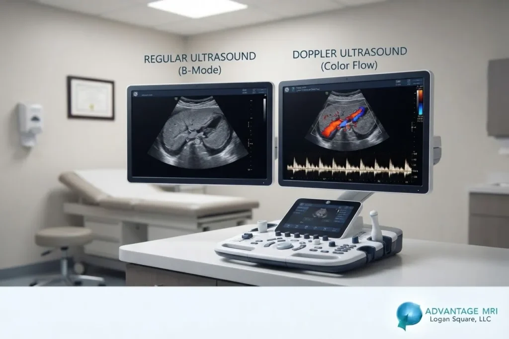
If your doctor mentioned a doppler test and you thought that sounded a lot like a regular ultrasound, you are not alone. A lot of patients hear both words in the same sentence and assume they mean the same thing. They do not. Regular ultrasound and doppler ultrasound both use sound waves to create pictures inside your body but they are built to answer different questions.
Regular ultrasound shows you what your organs and tissues look like. Doppler ultrasound shows you how your blood is moving through your arteries and veins. Once you understand that doppler vs ultrasound difference, most of the confusion clears up fast.
This article walks through what each test does, how they work, when your doctor might order one over the other and what you can expect if you come in for a doppler ultrasound at Advantage MRI in the Hermosa neighborhood of Chicago.

What Is Regular Ultrasound
Regular ultrasound, sometimes called diagnostic ultrasound or sonography, uses high frequency sound waves to build a picture of the organs and soft tissue inside your body. A technologist called a sonographer moves a small handheld device called a transducer across your skin, usually with a bit of gel to help the sound waves travel. The transducer sends sound waves into your body and those waves bounce off organs, muscles and tissue. The machine picks up the returning waves and turns them into an image on a screen.
This is the same basic technology used to look at a growing fetus during pregnancy, check the gallbladder, examine the liver or look at the thyroid. It shows structure. Shape, size, texture, whether something looks swollen or has a mass on it. What it does not show, at least not on its own, is how blood is flowing through the area.
Regular ultrasound does not use radiation like x rays do, which is part of why it is considered such a safe and widely used imaging tool. The American College of Radiology notes that ultrasound imaging carries no known risk from the sound waves themselves, which is one reason it is used so often in pregnancy and pediatric care.
Quick answer: Regular ultrasound uses sound waves to show the structure of organs and tissue. It does not measure blood flow speed or direction.
| Feature | Regular Ultrasound |
|---|---|
| What it shows | Organs, tissue structure, masses |
| Uses sound waves | Yes |
| Shows blood flow | No |
| Typical scan time | 15 to 30 minutes |
| Radiation | None |
| Common uses | Pregnancy, gallbladder, thyroid, abdomen |
For patients coming in for a standard scan, our ultrasound page explains what to expect step by step.
What Is Doppler Ultrasound
Doppler ultrasound is built on the same sound wave technology but it adds something regular ultrasound cannot do on its own. It measures the speed and direction of blood flow within your arteries and veins.
Here is the part that actually explains the name. The doppler effect is a change in pitch that happens when a sound source is moving toward you or away from you. Think of a siren on an ambulance. It sounds higher pitched as it comes toward you and lower pitched as it moves away. Doppler ultrasound uses that same principle, except instead of a siren, it is tracking sound waves bouncing off moving red blood cells.
When sound waves hit red blood cells that are moving toward the transducer, the frequency of the returning wave shifts higher. When blood is moving away, the frequency shifts lower. The machine reads that shift and translates it into information about how fast blood is moving and which direction it is going.
There are a few different doppler modes and each one is used for a slightly different job.
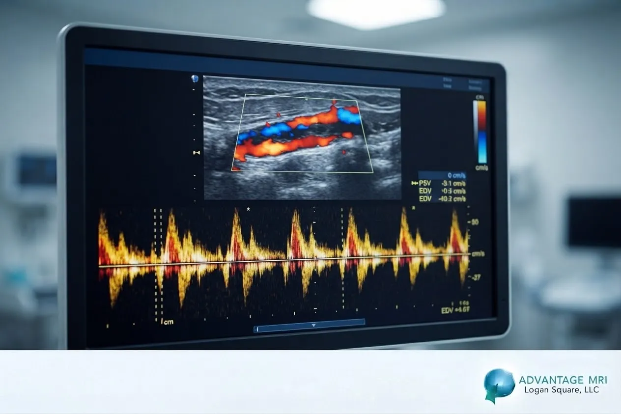
- Color doppler adds color to the image so you can see blood flow direction at a glance, usually red for flow toward the transducer and blue for flow away from it.
- Spectral doppler shows blood flow as a waveform or graph, which lets the radiologist study the speed and pattern of flow over time.
- Power doppler is more sensitive and picks up smaller or slower blood flow, which can be useful in smaller vessels.
- Transcranial doppler looks at blood flow in and around the brain, often through the skull and is sometimes used to check for issues related to stroke risk.
Quick answer: Doppler ultrasound uses the same sound wave imaging as regular ultrasound but adds the ability to measure the speed and direction of blood flow through blood vessels.
Doppler vs Ultrasound: The Differences
So what does this actually mean for you as a patient? Here is the short version. Regular ultrasound answers the question what does this look like. Doppler ultrasound answers the question how is blood moving through here.
A radiologist might order a regular ultrasound if there is a lump, unexplained pain or a need to check organ size. A doppler ultrasound gets ordered when there is concern about blood flow itself, things like a possible blood clot, narrowing in an artery or poor circulation in the legs.
| Feature | Regular Ultrasound | Doppler Ultrasound |
|---|---|---|
| Shows structure | Yes | Sometimes, depending on mode |
| Shows blood flow speed and direction | No | Yes |
| Used for | Organs, tissue, pregnancy | Blood clots, artery blockages, vein problems |
| Common areas checked | Abdomen, pelvis, thyroid | Legs, neck, arms, brain vessels |
| Time needed | 15 to 30 minutes | 20 to 45 minutes |
Both tests are noninvasive, meaning nothing goes inside your body and neither one uses radiation. That part is exactly the same. The difference comes down to what information the machine is set up to capture and translate.
Is Doppler Ultrasound the Same as Duplex Ultrasound
This one trips people up almost as often as doppler vs regular ultrasound. Duplex ultrasound is not a separate technology. It is a combination.
A duplex scan uses regular grayscale ultrasound to look at the structure of a blood vessel and doppler ultrasound at the same time to measure blood flow through that same vessel. So instead of choosing between seeing the vessel and measuring the flow, duplex ultrasound gives you both in one exam.
This is often what people are picturing when they ask about a doppler test for their legs. A lower extremity duplex ultrasound, for example, checks both the shape of the veins and how well blood is moving through them, which is useful for spotting a deep vein blood clot or valve problems that cause blood to pool.
Quick answer: Duplex ultrasound combines regular ultrasound imaging with doppler blood flow measurement in a single test.
When Doctors Order Doppler Instead of Regular Ultrasound
A doctor is not choosing between these tests at random. The choice usually comes down to your symptoms and what they need to rule in or rule out.
Doppler or duplex ultrasound tends to get ordered when there is leg swelling or pain that could point to a blood clot, when someone has cold or numb legs that might mean poor circulation, when there is a whooshing sound heard in the neck that could suggest a narrowed carotid artery or when someone with diabetes needs their circulation checked regularly.
Regular ultrasound is more likely to come into play for abdominal pain, an unexplained lump, monitoring a pregnancy or checking on an organ that showed something unusual on another test.
There is also overlap with heart imaging here. An echocardiogram actually uses a form of doppler technology to look at blood flow through the heart’s chambers and valves, which is a good example of how doppler shows up in more places than people expect.
What to Expect During a Doppler Ultrasound
Most patients are surprised by how simple the process actually is. There is no needle, no radiation and in most cases no fasting required, though your provider may give you specific instructions depending on the area being scanned.
Before the Scan
You will usually be asked to wear loose clothing so the area being examined is easy to reach. If the scan is on your legs, you might be asked to avoid tight socks or compression garments beforehand. Some abdominal doppler scans, like those checking blood flow to the kidneys, may require a short fasting period. Your scheduling team will let you know what applies to your specific exam.
During the Scan
You will lie down on an exam table and gel gets applied to the area being checked. The sonographer moves the transducer slowly over your skin. You might hear a whooshing or pulsing sound during the exam. That sound is actually the doppler signal itself and it is completely normal. Most doppler ultrasounds take between 20 and 45 minutes, depending on how many vessels or areas need to be checked.
After the Scan
There is no recovery time needed. You can drive yourself home and go back to your normal activities right away. A radiologist reviews the images and doppler readings and your results typically get sent to your referring doctor within a day or two.
This is general information and is not a substitute for medical advice from your own doctor. If you have specific symptoms or health concerns, talk to your physician about which imaging test is right for you.
Doppler Ultrasound in Chicago: Why Patients Choose Advantage MRI
Advantage MRI is located at 2008 N Pulaski Rd in the Hermosa neighborhood of Chicago and doppler and duplex ultrasound are part of our regular imaging services. Our center is ACR accredited, which means we meet the American College of Radiology’s standards for equipment, staffing and image quality.
Our radiology team is led by Dr. Gregory T. Goldstein, a board certified diagnostic radiologist with a background in orthopedic and neurological imaging. Dr. Goldstein founded MetisMD and created Connected RadiologySM, a model built around fast, accurate reporting and close coordination with referring physicians. That matters here because doppler results often need a quick turnaround, especially when a possible blood clot is involved.
We also offer on site scheduling that tends to move faster than a lot of larger hospital systems, plus an on site pharmacy and IV capability for patients who need it during their visit. If your doctor has referred you for a doppler or duplex ultrasound or you are trying to figure out which imaging service fits your symptoms, our MRI imaging and ultrasound pages break down what each service covers.
FAQ
Is doppler same as ultrasound?
No. Doppler ultrasound is a type of ultrasound that adds the ability to measure blood flow speed and direction. Regular ultrasound only shows structure, not how blood is moving.
What is the difference between ultrasound doppler and duplex?
Doppler ultrasound measures blood flow on its own. Duplex ultrasound combines regular grayscale ultrasound with doppler in the same exam, so you get a picture of the vessel structure and the blood flow at the same time.
Does doppler ultrasound hurt?
No. Doppler ultrasound is noninvasive and painless. You may feel light pressure from the transducer but there is no needle and no cutting involved.
How long does a doppler ultrasound take?
Most doppler ultrasound exams take between 20 and 45 minutes, depending on how many blood vessels or areas need to be checked.
Can doppler ultrasound detect a blockage?
Yes. Doppler ultrasound can detect narrowing or blockages in arteries by showing changes in blood flow speed and pattern at the site of the blockage.
Do I need to prepare for a doppler scan?
It depends on the area being examined. Some doppler exams need no prep at all, while others, like abdominal doppler studies, may require a short fasting period. Your imaging center will give you specific instructions ahead of time.
Is doppler ultrasound covered by insurance?
In most cases, yes, when it is ordered by a physician for a medical reason. Coverage details vary by insurance plan, so it is worth checking with your provider or contacting our office directly.
What conditions is doppler ultrasound used to check?
Doppler ultrasound is commonly used to check for blood clots, especially deep vein thrombosis in the legs, narrowing in the carotid arteries, poor circulation and blood flow problems related to diabetes.
Can regular ultrasound replace a doppler test?
Not for blood flow concerns. Regular ultrasound cannot measure blood flow speed or direction, so if a blood clot or circulation issue is suspected, doppler or duplex ultrasound is the appropriate test.
How fast are doppler ultrasound results available?
A radiologist typically reviews the images shortly after the exam and results are usually sent to your referring physician within one to two business days, sometimes faster for urgent cases.
Conclusion
The difference between doppler and regular ultrasound really comes down to one thing. Regular ultrasound shows you what a structure looks like. Doppler ultrasound shows you how blood is moving through it. Duplex ultrasound gives you both at once. None of these tests involve radiation, needles or downtime, which is part of why they get used so often for everything from pregnancy checkups to blood clot concerns.
If your doctor has recommended a doppler or ultrasound exam or you are not sure which one fits your symptoms, our team at Advantage MRI in Chicago can help walk you through it. For reliable, advanced diagnostic imaging you can trust, call Advantage MRI today or schedule your appointment online.
Contact Us
- Address: 2008 N Pulaski Rd, Chicago, IL 60639
- Phone: (773) 227 9777
- Email: info@advantagemri.org
Follow us on social media for the latest health tips, diagnostic imaging updates and clinic news: Facebook | Instagram | LinkedIn.