Health & Imaging
Transthoracic vs Transesophageal Echocardiogram: Know the Difference
Your doctor says you need an echocardiogram and then drops two terms on you that sound almost the same. Transthoracic. Transesophageal. Whats the actual difference and why does it matter which one you get.
Here is the short answer. A transthoracic echocardiogram is the standard heart ultrasound done from outside your chest. A transesophageal echocardiogram uses a small probe passed down your esophagus to get a closer clearer view of your heart valves and chambers. Both use sound waves. Both create images of your heart. But they are not interchangeable and your cardiologist picks one based on what they need to see.
This article breaks down the transthoracic echocardiogram vs transesophageal echocardiogram question in plain language, so you know what to expect before your test at Advantage MRI in Chicago.
What Is an Echocardiogram
An echocardiogram is an imaging test that shows the structure and function of your heart. No needles involved in the basic version, no radiation either. It relies on sound waves, the same basic technology used in obstetric ultrasound, just aimed at your heart instead.
How Echocardiography Uses Sound Waves
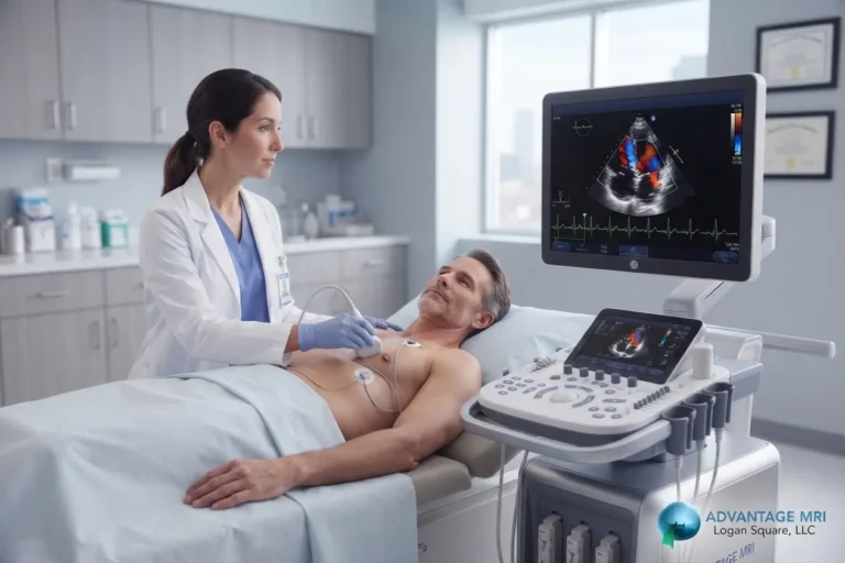
A handheld device called a transducer sends sound waves into your chest. Those waves bounce off your heart tissue, blood and valves, then bounce back to the transducer. A computer turns that returning signal into a moving image. Thats echocardiography in one sentence.
Doctors use this technique to measure how well your heart pumps, check your heart valve function, look at blood flow through the chambers and spot problems with the heart muscle itself. A version called Doppler echocardiography adds color and motion tracking so the sonographer can see blood flow direction and speed, which matters a lot when checking for a leaky or narrowed valve.
| What an Echocardiogram Checks | Why It Matters |
|---|---|
| How well your heart pumps | Shows the amount of blood pumped out with each beat, an indication of overall heart function |
| Heart valve function | Helps spot valve problems like leaking or narrowing before they get worse |
| Blood flow through the chambers | Doppler tracking shows blood flow direction and speed, useful for catching flow issues early |
| Structure of the heart muscle | Flags problems with the heart muscle itself that a physical exam alone cannot detect |
What Is a Transthoracic Echocardiogram (TTE)
A transthoracic echocardiogram is the version most people picture when they hear the word echocardiogram. Its the simplest type of echocardiography and the one used first in almost every case.
How TTE Test Works
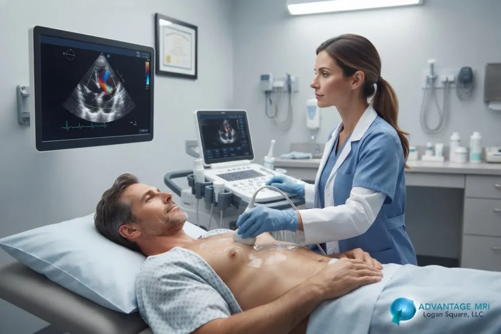
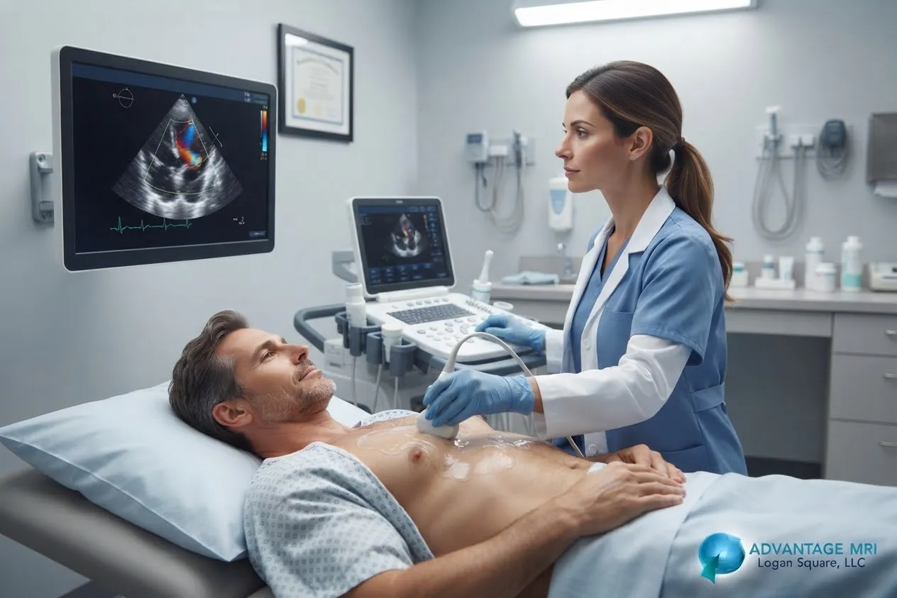
You lie on a table or bed, usually on your left side. A sonographer applies gel to your chest and moves the transducer across different spots to capture the heart from several angles. The sound waves pass through your skin and chest wall to reach the heart underneath. No sedation is needed, no fasting required in most cases and the whole thing usually takes under an hour.
A transthoracic echo is painless, noninvasive and can be performed on an outpatient basis or as part of a broader cardiac workup. Once its done you can typically resume your usual diet and activities right away.
What TTE Shows About Heart Valve Function
A TTE gives your cardiologist an actual picture of heart structures including the four chambers and four valves. It can measure the amount of blood pumped out with each heartbeat, an indication of how well your heart is functioning overall. It can also flag early signs of heart valve disease, an enlarged chamber or fluid around the heart.
For a lot of people, a transthoracic echocardiogram answers every question the doctor needed answered. It only moves to something more invasive when the images from outside the chest are not clear enough or when a specific structure needs a closer look.
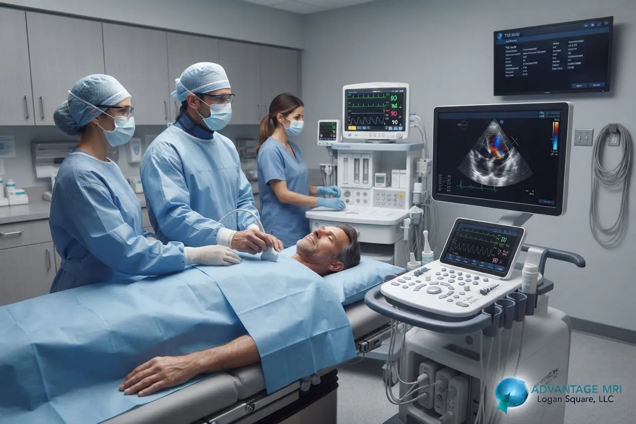
What Is a Transesophageal Echocardiogram (TEE)
A transesophageal echocardiogram or TEE, takes a different path. Instead of sending sound waves through the chest wall, the TEE probe goes down your esophagus, which sits directly behind your heart. Since the esophagus and heart are so close together, the probe is much closer to the heart than a handheld device pressed against your skin ever could be. That closeness is the whole point.
Why TEE Needs Sedation
Youll get a numbing spray on the back of your throat and usually a mild sedative through an IV before the TEE probe goes in. That combination keeps you relaxed and comfortable while the thin flexible tube with a small transducer on the end is guided down. Most people dont remember much of the procedure once the sedation kicks in.
Because of the sedation, you cannot drive yourself home afterward. Someone else needs to take you. Youll also need to fast for a period beforehand, similar to prep for an endoscopy, since the probe passes through the same area used for swallowing.
TEE and Valve Replacement Planning
A transesophageal echocardiogram gives a much sharper image of the heart valves, especially the mitral valve, which sits toward the back of the heart and can be hard to see clearly on a standard chest ultrasound. This is why TEE gets used so often before valve replacement surgery, when a surgeon needs precise detail on valve structure before going in. It is also the preferred test for spotting infections like endocarditis, checking for blood clots before a procedure like cardioversion and evaluating certain congenital heart conditions.
Transthoracic and Transesophageal: Differences
Both tests use sound waves and both create images of your heart. That is where the similarities mostly end.
| TTE | TEE | |
|---|---|---|
| Probe Location | Transducer moved across the outside of the chest | Probe passed down the esophagus, close behind the heart |
| Sedation Needed | No sedation required | Numbing throat spray plus a mild IV sedative |
| Image Clarity | Clear overall picture but some views can be blocked by skin, muscle or lung tissue | Sharper closer images, especially of the mitral valve and back heart structures |
| Prep Required | No fasting, no special prep in most cases | Fasting beforehand and a driver arranged for after sedation |
| Typical Duration | About thirty to sixty minutes | Longer visit due to sedation and recovery time before going home |
| Common Use Case | First test ordered for most heart evaluations | Valve replacement planning, suspected valve infection or checking for blood clots |
Accuracy Differences
Because the TEE probe sits closer to the heart with fewer layers of tissue in the way, it produces clearer detailed images, particularly of the back structures like the mitral valve and the upper heart chambers. A TTE image passes through skin, muscle and sometimes lung tissue or ribs, which can blur or block certain views, especially in patients with lung disease or a larger body frame.
Comfort and Recovery Differences
A TTE requires no sedation, no fasting and no downtime. You walk in, get scanned and walk out. A TEE is more involved. You need sedation, a driver, a period of fasting before the test and some recovery time afterward since your throat will be numb and youll want that to wear off before eating or drinking again.
Neither test is something to be scared of. The truth is most people tolerate both just fine and your care team will walk you through exactly what to expect before anything starts.
Types of Echocardiogram Beyond TTE and TEE
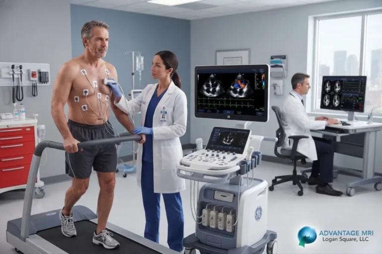
Stress Echocardiogram Explained
A stress echocardiogram checks how your heart handles exertion. You either walk on a treadmill or get a medication injected that mimics the effect of exercise on your heart rate. Images are taken before and after to compare how the heart moves under stress versus at rest. This version helps diagnose coronary artery disease when doctors suspect the heart isnt getting enough blood flow during activity.
There is also intracardiac echocardiography, a more specialized version used during certain cardiac procedures, though thats far less common and typically only relevant in a hospital procedural setting rather than routine outpatient testing.
Which One Does Your Doctor Choose
Your cardiologist starts with a transthoracic echocardiogram in almost every situation. Its noninvasive, quick and gives a solid overall picture of heart health. If those images answer the clinical question, thats the end of it.
A transesophageal echocardiogram vs transthoracic decision usually comes down to one thing: does the doctor need a clearer or closer look than the TTE could provide. That might be because the TTE images were unclear, because theyre checking for a blood clot before a heart rhythm procedure, because theyre planning valve replacement surgery or because theyre trying to rule out an infection on a heart valve.
This isnt something you need to figure out on your own. Your referring physician and the imaging team communicate about which test fits your specific situation and youll be told clearly which one youre scheduled for and why.
What Happens During Your Visit at Advantage MRI
At Advantage MRI in the Hermosa neighborhood of Chicago, echocardiogram testing is handled by a team that understands both the technical side and the human side of the experience. Our echocardiogram services are performed with attention to comfort and clear communication, so you know what each step involves before it happens.
Diagnostic imaging at Advantage MRI is reviewed with board certified radiologist oversight, including Dr. Gregory T. Goldstein, MD, a board certified diagnostic radiologist with a background in orthopedic and neurological imaging. That level of review matters when precision affects what your cardiologist decides next.
Beyond echocardiogram testing, Advantage MRI also offers ultrasound imaging, EKG testing to check your hearts electrical activity and Holter monitor monitoring for patients whose symptoms come and go. If your workup involves nerve related symptoms, we also provide nerve conduction study and EMG testing services, along with MRI imaging and digital x ray for a full range of diagnostic needs.
Want to learn more about our team first? Visit our about us page.
This article is for general information only and is not a substitute for professional medical advice. Talk with your doctor about which echocardiogram test is right for your specific heart health needs.
Frequently Asked Questions About Transthoracic Echocardiogram vs Transesophageal Echocardiogram
What is the main difference between TTE and TEE?
A TTE uses a transducer moved across your chest from outside the body, while a TEE uses a probe passed down your esophagus to get a closer image of the heart. TTE needs no sedation, TEE does.
Does a TEE echocardiogram hurt?
Most people describe mild discomfort during probe insertion, similar to swallowing something slightly larger than usual. The numbing spray and sedation keep this manageable and many patients dont recall much of the procedure afterward.
How long does a transthoracic echocardiogram take?
A standard TTE usually takes between thirty and sixty minutes, depending on how many views the sonographer needs to capture a complete picture of the heart.
Is sedation always needed for TEE?
Yes, in nearly all cases. A mild IV sedative along with a numbing throat spray is standard for a TEE to keep you comfortable while the probe is guided into position.
Can a TTE miss things a TEE catches?
Sometimes. Certain structures, especially the back of the heart and the mitral valve, are harder to see clearly through the chest wall. When those areas need closer evaluation, a TEE fills that gap.
How do I prepare for a TEE test?
Youll typically need to fast for several hours beforehand and arrange for someone to drive you home after the sedation wears off. Your care team will give you specific instructions after the procedure is scheduled.
Is an echocardiogram the same as an EKG?
No. An EKG records the electrical activity of your heart through sensors on your skin, while an echocardiogram creates an actual image of your hearts structure using sound waves. Doctors often order both together for a fuller picture of heart health.
Who typically needs a TEE instead of a TTE?
Patients being evaluated for valve replacement surgery, suspected heart valve infection, blood clots before certain heart procedures or unclear TTE images are the most common candidates for a TEE.
How soon do I get echocardiogram results?
Results are reviewed by a radiologist or cardiologist and typically shared with your referring physician within a few business days, though timing can vary based on the reason for the test.
Does insurance cover echocardiogram testing?
Most insurance plans cover echocardiogram testing when its ordered by a physician for a medical reason. Coverage details vary by plan, so its worth confirming your specific benefits before your appointment.
Conclusion
A transthoracic echocardiogram and a transesophageal echocardiogram both exist to answer the same basic question: how is your heart doing. TTE handles that job for most people, quickly and without any invasive steps. TEE steps in when a closer sharper view of the valves and chambers is needed, particularly ahead of valve replacement or when checking for infection or clots. Neither test should feel like a mystery once you understand what each one involves and why your doctor chose it.
Contact Us
- Address: 2008 N Pulaski Rd, Chicago, IL 60639
- Phone: (773) 227 9777
- Website: advantagemri.org
For reliable advanced diagnostic imaging you can trust, call Advantage MRI today or schedule your appointment online. You can also reach out through our contact us page with any questions.
Follow us on social media for the latest health tips, diagnostic imaging updates and clinic news: Facebook | Instagram | LinkedIn.