Health & Imaging
PET Scan vs CT Scan vs MRI: A Plain-English Guide to the Three Main Scan Types
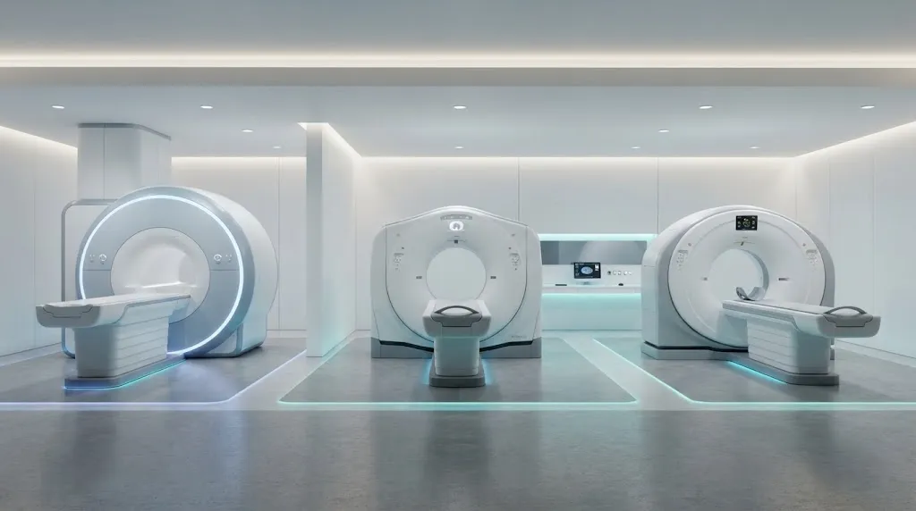
Quick answer: The biggest difference is what each scan shows. A CT scan uses X-rays to show the body’s structure quickly, especially bone, lungs and bleeding.When comparing pet scan vs ct scan vs mri, an MRI uses magnets and radio waves to show soft tissues, such as the brain, spinal cord, joints and organs, in fine detail without radiation.
A PET scan uses a small amount of radioactive tracer to show how tissues are working, such as how actively cells are using sugar, which is especially useful in cancer, brain disorders and some heart conditions. PET is often combined with CT or MRI in a single exam so doctors see both activity and anatomy.
Doctors often say CT and MRI show what things look like, while PET shows what things are doing. This guide explains each test, compares them side by side, and covers safety, preparation and when each one is usually used.
Need to Schedule a Diagnostic Appointment?
Whether you need an MRI, ultrasound, X-ray, or another diagnostic service, Advantage MRI is here to help you take the next step in your healthcare journey.
Submit your appointment request online, and our team will contact you to confirm your visit.
Book Your Appointment →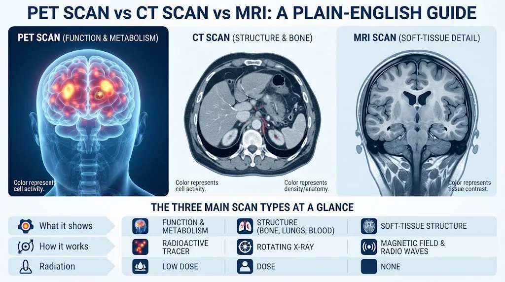
PET vs CT vs MRI at a Glance
| Feature | PET scan | CT scan | MRI |
|---|---|---|---|
| What it shows | Function and metabolism (cell activity) | Structure, especially bone, lungs and blood | Soft-tissue structure in high detail |
| How it works | Detects a radioactive tracer injected into a vein | Rotating X-ray beam and computer processing | Strong magnetic field and radio waves |
| Ionizing radiation | Yes (from the tracer, plus CT if combined) | Yes | No |
| Typical scan time | Scan often 20-30 min, total visit often 2 hours or more | Seconds to a few minutes | Often 20-60 minutes |
| Preparation | Usually fasting and blood sugar control | Often little; sometimes fasting for contrast | Metal screening; sometimes fasting |
| Common uses | Cancer staging and treatment response, some dementia and epilepsy workups, heart muscle viability | Trauma, chest and abdominal emergencies, lung nodules, kidney stones | Brain, spine, joints, ligaments, many organ problems |
| Relative cost | Usually highest | Usually lowest of the three | Usually between CT and PET |
| Availability | Fewer centers | Very widely available | Widely available |
What Is a CT Scan?
Computed tomography (CT) takes many X-ray images from different angles as the scanner rotates around you. A computer combines them into thin cross-sectional “slices” and 3D reconstructions.
Strengths:
- Very fast, which matters in emergencies and for patients who can’t stay still long
- Excellent for bone, lungs and detecting bleeding
- Widely available, including 24/7 in emergency departments
- Generally less expensive than MRI or PET
Limitations:
- Uses ionizing radiation
- Less soft-tissue contrast than MRI, for example in the spinal cord, ligaments or subtle brain changes
- Iodine-based contrast, when needed, isn’t suitable for everyone
Common CT uses: head injury, suspected bleeding in the brain, chest pain workups that need CT angiography, lung nodules, kidney stones, appendicitis and other abdominal emergencies, and complex fractures.
What Is an MRI?
Magnetic resonance imaging (MRI) places you in a strong magnetic field. Radio wave pulses briefly disturb hydrogen atoms in your body, and the signals they give off as they settle are turned into images. Because different tissues give off different signals, MRI shows soft tissue in remarkable detail.
Strengths:
- No ionizing radiation
- Outstanding soft-tissue contrast for the brain, spinal cord, muscles, tendons, ligaments, cartilage and many organs
- Many specialized sequences for blood flow, diffusion and inflammation
Limitations:
- Takes longer than CT
- Loud and enclosed, which can be hard for people with claustrophobia
- Some implants and metal objects are unsafe or require special protocols
- Usually costs more than CT
Common MRI uses: brain and spinal cord conditions, back and neck pain with nerve symptoms, sports injuries, joint problems, certain liver, pelvic and prostate conditions, and many soft-tissue masses. For a closer look at how MRI and CT compare across the whole body, read our MRI vs CT scan pillar guide.
What Is a PET Scan?
Positron emission tomography (PET) is a nuclear medicine test. Before the scan, a small amount of a radioactive tracer is injected into a vein. The most common tracer is FDG, a form of glucose (sugar). Cells that are very active, such as many cancer cells, take up more glucose than surrounding tissue, so they “light up” on the scan.
After the injection, you rest quietly for roughly an hour while the tracer spreads through your body. Then you lie on the scanner table while the PET camera detects the tracer’s signals.
Strengths:
- Shows metabolic activity, which can reveal disease before structural changes are visible
- Helps doctors see whether cancer has spread and whether treatment is working
- Specialized tracers can look at specific targets, such as amyloid in the brain or certain prostate cancer markers
Limitations:
- Uses radiation from the tracer (and from the CT portion if combined)
- Lower spatial detail on its own, which is why it is usually paired with CT or MRI
- Requires careful preparation, including blood sugar control
- Inflammation and infection can also take up tracer, which can cause false positives
- Usually the most expensive of the three and less widely available
Common PET uses: cancer diagnosis, staging and monitoring; evaluating some causes of dementia; locating seizure sources in epilepsy; and assessing heart muscle viability or blood flow.
Why PET Is Usually Combined With CT or MRI
A PET image on its own shows bright spots of activity but limited anatomical detail. Combining it with CT or MRI tells doctors exactly where the activity is.
- PET/CT is the most common combination. The CT portion maps anatomy and helps correct the PET images. Both scans are done on the same machine in one session.
- PET/MRI combines PET with MRI’s soft-tissue detail and lower radiation from the anatomical part. It is available at fewer centers and is used in selected cases, such as some brain, pelvic and pediatric exams.
PET Scan vs MRI: The Most Common Comparison
People often ask whether they should have a PET scan or an MRI. These tests answer different questions, so one is not simply better than the other.
| Question your doctor is asking | Test that usually helps more |
|---|---|
| Is there a structural problem in the brain or spine? | MRI |
| Has a known cancer spread elsewhere in the body? | PET/CT |
| Is a cancer responding to treatment? | Often PET/CT, sometimes MRI or CT |
| Is a knee, shoulder or ankle ligament torn? | MRI |
| What type of dementia might this be? | MRI first, PET in selected cases |
| Where in the brain are seizures starting? | MRI first, PET when MRI is unclear |
| Is a liver lesion benign or concerning? | Often MRI |
In many cases, doctors use both tests at different stages. For example, MRI may find a brain tumor, and PET may later help evaluate treatment response.
PET vs CT: What’s the Difference?
CT shows shape and density. PET shows activity. A CT scan can show that a lymph node is enlarged, but it can’t always tell whether the enlargement comes from cancer or from inflammation. PET can show whether that node is metabolically active, which adds another clue. Because each test fills in what the other misses, PET and CT are frequently performed together.
Safety Comparison
Radiation
- MRI: no ionizing radiation.
- CT: uses ionizing radiation. The dose depends on the body part and the protocol.
- PET: the tracer is radioactive, and a PET/CT adds CT radiation. The tracer decays quickly, and most of it leaves the body within hours.
For a medically justified scan, doctors judge that the benefit of the information outweighs the small radiation risk. If you’ve had many scans, tell your doctor.
Pregnancy and Breastfeeding
Tell your care team if you are or might be pregnant, or if you are breastfeeding. MRI without contrast is generally preferred when imaging is needed in pregnancy. PET and CT are used in pregnancy only when the benefit clearly outweighs the risk. After PET, breastfeeding mothers may receive specific instructions about timing and close contact.
Contrast and Tracers
- CT contrast is iodine-based.
- MRI contrast is gadolinium-based.
- PET tracers are given in very small amounts and rarely cause allergic reactions.
People with kidney disease or previous contrast reactions should tell the imaging team in advance.
Metal and Implants
MRI requires the most careful screening because of the strong magnet. Pacemakers, defibrillators, cochlear implants, neurostimulators, certain aneurysm clips and metal fragments must be checked. CT and PET generally have fewer restrictions related to metal, although metal can create image artifacts.
How to Prepare for Each Scan
Preparing for a CT
- Remove metal from the area being scanned.
- If contrast is used, you may be asked to avoid eating for a few hours.
- Tell staff about kidney problems, diabetes medications and previous contrast reactions.
Preparing for an MRI
- Complete the metal and implant safety questionnaire honestly.
- Leave jewelry, watches, cards and hairpins at home if possible.
- Fasting depends on the body part and whether contrast is used. See our guide on eating before an MRI.
- Mention claustrophobia ahead of time.
Preparing for a PET Scan
- You will usually be asked to fast for several hours before the scan.
- Avoid strenuous exercise the day before, because muscle activity can affect tracer uptake.
- If you have diabetes, you will get specific instructions about food and medication, since high blood sugar can affect the results.
- Plan for a longer appointment that includes the tracer uptake time.
- Drink fluids afterward, if allowed, to help flush the tracer.
Always follow the specific instructions from the facility performing your exam, since protocols vary.
How Long Does Each Scan Take?
- CT: The scan takes seconds to minutes. Many visits are done in under 30 minutes, longer if oral contrast is needed.
- MRI: Usually 20-60 minutes in the scanner, depending on the body part.
- PET/CT: The scan portion is often 20-30 minutes, but the visit commonly lasts two hours or more because of check-in, injection and uptake time.
Cost Differences
Costs vary widely depending on location, facility type, body part, contrast use and insurance. As a general pattern:
- CT tends to be the least expensive.
- MRI usually costs more than CT.
- PET/CT is usually the most expensive.
Independent outpatient imaging centers often charge less than hospital outpatient departments for the same test. Insurers commonly require prior authorization for MRI and PET. Ask for a written estimate if you are paying out of pocket.
How the Three Scans Work Together: Real-World Examples
Patients often have more than one of these scans, each at a different stage. These simplified examples show typical patterns. Your own care plan may differ.
Example 1: A lung nodule. A chest CT done for another reason shows a small spot on the lung. The doctor may order a follow-up CT in a few months to see if it changes. If the nodule is larger or looks suspicious, a PET/CT may be used to see whether it is metabolically active and whether anything has spread. A biopsy is often needed for a definite answer.
Example 2: New neurological symptoms. A person develops numbness and vision changes. A brain and spine MRI shows lesions consistent with multiple sclerosis. CT and PET usually aren’t needed in this situation because MRI answers the question best.
Example 3: A diagnosed lymphoma. After diagnosis, PET/CT is commonly used to stage the disease, meaning to find all the areas involved. Another PET/CT during or after treatment can show whether the active areas have responded.
Example 4: A knee injury. An X-ray is done first to rule out a fracture. If pain and instability continue, an MRI shows ligaments, menisci and cartilage. PET and CT are rarely part of this pathway.
Example 5: Memory loss. An MRI looks for structural causes, such as strokes, tumors or fluid buildup, and shows patterns of shrinkage. In selected cases, a specialist may add a PET scan that looks at brain metabolism or amyloid deposits to help clarify the type of dementia.
The pattern is clear: CT answers fast structural questions, MRI answers detailed soft-tissue questions, and PET answers questions about activity. Choosing the right order saves time, cost and radiation.
Which Scan Will Your Doctor Order?
Your doctor chooses based on:
- The clinical question. Structure, soft-tissue detail or function?
- Urgency. Emergencies often favor CT for speed.
- The body part. Joints and the spinal cord favor MRI; lungs and bone often favor CT.
- Your safety profile. Implants, kidney function, pregnancy and radiation history.
- Evidence-based guidelines. Many clinicians use the American College of Radiology Appropriateness Criteria.
- Insurance requirements. Some plans require one test before another. For example, an X-ray is often required before an MRI for certain joint problems. Our MRI vs X-ray guide explains why.
For brain-specific decisions, read our guide on which is better for the brain, MRI or CT. If you’re considering a scan with no symptoms at all, our article on full-body MRI screening covers the evidence and limitations.
Imaging Services at Advantage MRI in Chicago
Advantage MRI is an outpatient diagnostic center at 2008 N Pulaski Rd, Chicago, IL 60639. We provide MRI imaging, digital X-ray, ultrasound and Doppler studies, echocardiograms, EKGs, Holter monitoring, EMG and nerve conduction studies. We do not perform PET or CT scans. If your doctor orders one of those, it is usually done at a hospital or a specialized imaging center.
If MRI is the right test for you, our team handles safety screening, scheduling and reporting back to your referring physician. We welcome insured, out-of-network and self-pay patients.
Frequently Asked Questions About Pet Scan VS CT Scan VS MRI
Is a PET scan more accurate than an MRI?
They measure different things. PET shows tissue activity, while MRI shows soft-tissue structure in detail. Accuracy depends on the question being asked. Many patients benefit from both at different times.
Can a PET scan detect cancer that an MRI misses?
Sometimes. PET can show metabolically active cancer spread in areas that look normal in size on other scans. MRI can detect some cancers that PET shows poorly, such as small brain lesions. They are often complementary.
Which scan has the most radiation?
MRI has none. Between CT and PET/CT, the combined PET/CT exam usually involves more radiation than a single CT of one body region, because it includes both the tracer and a CT component.
Why is a PET scan usually done with a CT scan?
PET shows activity but limited anatomical detail. The CT portion shows exactly where that activity is and improves image accuracy.
Is an MRI safer than a CT scan?
MRI avoids ionizing radiation, which is a safety advantage. However, MRI isn’t safe for some implants and metal objects. The safest test is the one that best answers your doctor’s question with the lowest overall risk for you.
How long after a PET scan am I radioactive?
The commonly used tracer decays quickly. Facilities usually give simple precautions for the rest of the day, such as limiting close contact with infants and pregnant people for a few hours. Follow the instructions you receive.
Do I need to fast before all three scans?
PET usually requires fasting. CT and MRI may or may not, depending on the body part and whether contrast is used. Always follow your facility’s instructions.
Conclusion
PET, CT and MRI each answer a different question. CT gives fast structural pictures and is the workhorse of emergency imaging. MRI provides detailed soft-tissue images without radiation. PET shows how tissues are functioning and is especially valuable in cancer care and some brain and heart evaluations. Many patients end up with more than one type over the course of their care, and that is often by design.
Has your doctor ordered an MRI in Chicago? Book your MRI appointment with Advantage MRI or call (773) 227-9777. Open Monday-Friday 9:00 AM-5:00 PM and Saturday 9:00 AM-1:00 PM. Follow us on Facebook and Instagram.