Health & Imaging
Venous Doppler Ultrasound: DVT, Leg Pain and Blood-Flow Testing Explained
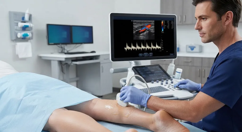
Quick answer: A venous Doppler ultrasound is a painless, radiation-free scan that checks how blood flows through your veins, most often in the legs. It is the main test doctors use to look for deep vein thrombosis (DVT), a blood clot in a deep vein. It can also evaluate leg swelling, varicose veins and leaky vein valves (venous insufficiency). Most leg studies take about 30 to 60 minutes and need no special preparation.
If you have sudden shortness of breath, chest pain, or are coughing up blood, especially with a swollen or painful leg, call 911. A clot can travel to the lungs (pulmonary embolism), which is an emergency.
This guide explains how a venous Doppler works, why it’s ordered, what happens during the exam and what results usually mean. We also explain how it differs from arterial, renal and liver Doppler studies, since those are often confused.
Need to Schedule a Diagnostic Appointment?
Whether you need an MRI, ultrasound, X-ray, or another diagnostic service, Advantage MRI is here to help you take the next step in your healthcare journey.
Submit your appointment request online, and our team will contact you to confirm your visit.
Book Your Appointment →What Is a Venous Doppler Ultrasound?
A standard ultrasound uses high-frequency sound waves to make images of structures inside the body. Doppler is an ultrasound technique that measures the movement of blood. When sound waves bounce off moving red blood cells, their pitch changes. The ultrasound machine turns that change into color maps and waveforms that show the speed and direction of blood flow.
A venous Doppler focuses on veins, the vessels that carry blood back to the heart. Most venous studies combine three techniques, often called a duplex or compression study:
- Grayscale (B-mode) imaging to see the vein walls and anything inside the vein
- Compression, where the sonographer gently presses on the vein with the probe; a healthy vein collapses, while a vein with a clot usually does not
- Color and spectral Doppler to confirm and measure blood flow
For a fuller explanation of the technology, see our article on Doppler vs standard ultrasound vs duplex.
Why Would a Doctor Order a Venous Doppler?
To Check for Deep Vein Thrombosis (DVT)
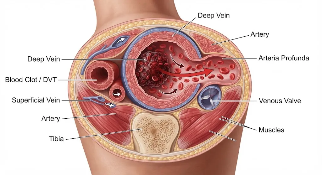
DVT is a blood clot in a deep vein, usually in the leg or pelvis. Common symptoms include:
- Swelling in one leg (sometimes both)
- Pain or tenderness, often in the calf or thigh
- Warmth or redness of the skin
- A feeling of heaviness or cramping
Some DVTs cause no symptoms at all. Risk factors include recent surgery or hospitalization, long periods of immobility (such as long flights or bed rest), cancer, pregnancy and the weeks after delivery, estrogen-containing birth control or hormone therapy, obesity, smoking, a previous clot, and inherited clotting disorders.
Doctors often combine a clinical scoring system (such as the Wells score) and a D-dimer blood test to decide how likely DVT is. When DVT is suspected, venous ultrasound is the standard imaging test.
To Evaluate Chronic Venous Insufficiency
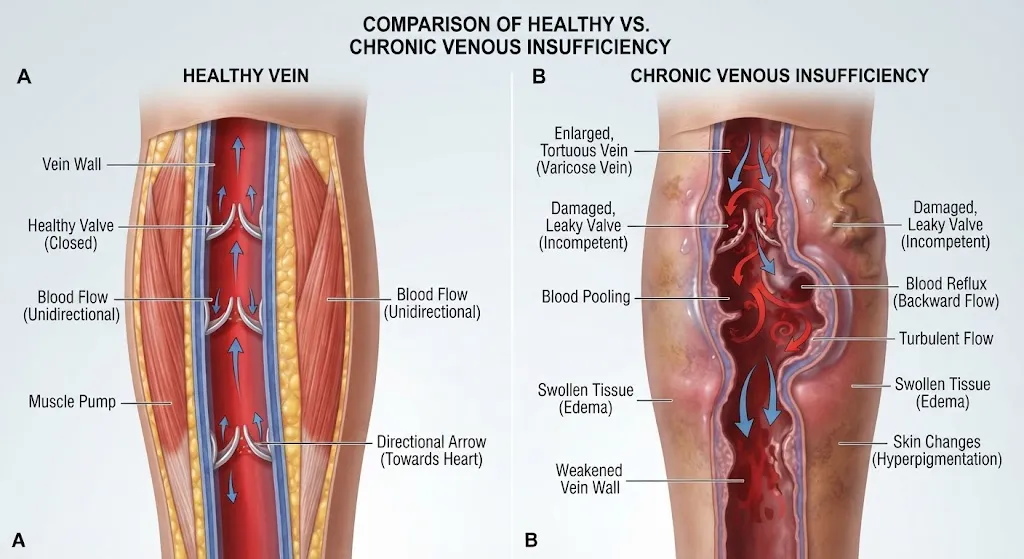
Veins have one-way valves that stop blood from flowing backward. When these valves weaken, blood pools in the legs. Signs include:
- Aching, tired or heavy legs, often worse at the end of the day
- Ankle swelling
- Varicose veins
- Skin darkening or thickening near the ankles
- Slow-healing sores (venous ulcers)
A venous reflux study is a specialized venous Doppler done to map which valves are leaking. It’s commonly ordered before vein treatment procedures.
Other Reasons
- Unexplained leg or arm swelling
- Follow-up of a known clot during treatment
- Checking veins before placing dialysis access or catheters
- Superficial thrombophlebitis (a clot in a surface vein) to make sure it hasn’t extended into deep veins
Doppler Ultrasound of the Legs: Veins vs Arteries
People searching for a “Doppler ultrasound of the legs” may be referring to one of two different tests:
| Venous Doppler | Arterial Doppler | |
|---|---|---|
| Vessels checked | Veins (blood returning to the heart) | Arteries (blood flowing from the heart) |
| Main conditions | DVT, venous insufficiency, varicose veins | Peripheral artery disease (PAD), narrowing or blockage |
| Typical symptoms | Swelling, calf pain, heaviness, skin changes | Leg pain while walking that eases with rest (claudication), cold feet, slow-healing toe or foot wounds |
| Often paired with | D-dimer blood test | Ankle-brachial index (ABI) blood pressure test |
What Is an Arterial Doppler Ultrasound?
An arterial Doppler measures blood flow through the arteries. It is used to detect peripheral artery disease, which happens when fatty plaque narrows the arteries, often in the legs. The study may be combined with an ankle-brachial index (ABI), which compares blood pressure in the ankles to the arms. PAD shares risk factors with heart disease, including smoking, diabetes, high blood pressure and high cholesterol.
Arterial Doppler is also used for the neck arteries to assess stroke risk. Our guide to carotid Doppler ultrasound covers that test.
Other Doppler Ultrasounds: Renal and Liver
Doppler isn’t limited to the legs.
Renal Doppler Ultrasound
A renal Doppler looks at blood flow in the kidneys and the arteries supplying them. Doctors may order it to:
- Look for renal artery stenosis (narrowing), which can cause hard-to-control high blood pressure
- Evaluate kidney blood flow in people with kidney disease
- Check a transplanted kidney
Preparation often includes fasting for several hours to reduce bowel gas, which can block the view. Your facility will tell you exactly what to do.
Doppler Ultrasound for the Liver
A liver (hepatic) Doppler checks blood flow through the portal vein, hepatic veins and hepatic artery. It can help evaluate:
- Portal hypertension, often related to cirrhosis
- Clots in the portal or hepatic veins
- Blood flow after liver procedures or transplantation
Fasting for several hours beforehand is commonly required.
How to Prepare for a Venous Doppler Ultrasound
For a standard leg or arm venous study, preparation is simple:
- No fasting is usually needed for limb studies.
- Wear loose, comfortable clothing. You may be asked to change into a gown or shorts.
- Take your usual medications unless your doctor tells you otherwise.
- Bring your order, insurance card and ID, plus any prior ultrasound reports.
- Tell the sonographer about recent surgeries, known clots, or areas that are especially painful.
For abdominal Doppler studies, such as renal or liver, follow fasting instructions carefully.
What Happens During the Exam?
- Positioning. You’ll usually lie on your back with your head slightly raised. For some parts of the exam, you may lie on your side, or your leg may be turned outward with the knee slightly bent. For venous reflux studies, part of the exam may be done standing.
- Gel. Warm, water-based gel is applied to your skin to help sound waves travel.
- Scanning. The sonographer moves a handheld probe (transducer) along the vein, typically from the groin down to the calf for a leg study.
- Compression. The sonographer presses gently every few centimeters to see if the vein collapses normally. If an area is tender, let them know. They can adjust.
- Doppler sounds. You may hear a whooshing sound from the machine. That’s the sound of blood flowing and is completely normal.
- Maneuvers. You might be asked to take a deep breath, hold it, or squeeze or relax your calf so the sonographer can check how blood responds.
- Finishing. The gel is wiped off, and you can return to normal activities right away.
The exam is generally painless. Some people with a tender, swollen leg notice brief discomfort during compression.
How Long Does a Venous Doppler Take?
- One leg: often about 30 minutes
- Both legs: often 45 to 60 minutes
- Venous reflux mapping: may take longer because it checks valve function in more detail
Allow extra time for check-in and paperwork.
What Do Venous Doppler Results Mean?
A radiologist or qualified physician reviews the images and sends a report to your doctor. Common findings include:
| Finding | What it generally means |
|---|---|
| Normal / no DVT | Veins compress fully and blood flows normally. No clot seen in the veins examined. |
| Acute DVT | A recent clot is present. The vein doesn’t compress and may be enlarged. Usually treated with blood thinners. |
| Chronic changes | Signs of an older clot or scarring of the vein wall. |
| Superficial thrombophlebitis | A clot in a surface vein. Doctors check whether it is close to a deep vein. |
| Venous reflux | Blood flows backward through leaky valves, consistent with venous insufficiency. |
| Other findings | Sometimes ultrasound shows a non-vein cause for symptoms, such as a Baker’s cyst behind the knee or a muscle injury. |
Only your doctor can explain how your results fit with your symptoms and history. If a DVT is found, treatment usually starts promptly, often with anticoagulant medication. Follow your doctor’s instructions closely.
Venous Doppler of the Arms
Clots can also form in the arm veins, though less often than in the legs. Upper-extremity DVT is more common in people who have a central line, PICC line, pacemaker wires or dialysis access, and sometimes in athletes who make repetitive overhead movements. Symptoms include arm or hand swelling, a heavy or aching arm, and visible veins across the shoulder or chest.
An arm venous Doppler follows the same principles as a leg study. The sonographer checks the veins from the neck and collarbone area down the arm, using compression where possible and Doppler where bone blocks compression, such as under the collarbone. Preparation is the same: no fasting, comfortable clothing, and a list of any lines or devices you have.
What Happens If a DVT Is Found?
If your scan shows a deep vein clot, your doctor will usually contact you quickly. Treatment decisions depend on where the clot is, what caused it, your bleeding risk and your overall health. Common steps include:
- Anticoagulant medication (“blood thinners”). These don’t dissolve the existing clot directly. They stop it from growing and lower the chance of new clots while your body gradually breaks the clot down. Treatment often lasts at least three months, sometimes longer.
- Monitoring for pulmonary embolism. Your doctor will explain warning signs, such as sudden shortness of breath, chest pain that worsens with breathing, a rapid heartbeat or coughing up blood.
- Compression stockings. Some patients use them to help with swelling and discomfort, based on their doctor’s advice.
- Follow-up ultrasound. A repeat scan may be ordered to check the clot, especially if it was in a calf vein and treatment wasn’t started right away.
- Looking for a cause. If there was no clear trigger, your doctor may consider further testing.
In uncommon, severe cases, procedures to remove or break up the clot may be considered by a specialist.
Lowering Your Risk of Leg Clots
Some risk factors can’t be changed, but these everyday habits can help, especially during travel or recovery:
- Move your legs regularly on long flights or drives; walk the aisle or stop every couple of hours when possible.
- Flex and point your feet and squeeze your calf muscles while sitting.
- Stay hydrated.
- Get up and walk as soon as you’re cleared after surgery, and follow any clot-prevention plan your surgical team gives you.
- Talk to your doctor about your clot risk before starting estrogen-containing medications, especially if you’ve had a clot before or have a family history.
- If you smoke, ask about support to quit.
How Accurate Is a Venous Doppler?
Compression ultrasound is very accurate for detecting clots in the larger veins of the thigh and behind the knee. It is somewhat less reliable for small calf veins and veins deep in the pelvis, where body size and bowel gas can limit visibility. If your first ultrasound is negative but suspicion remains high, your doctor may repeat the scan after a few days or order a different test.
Is a Venous Doppler Safe?
Yes. Doppler ultrasound uses sound waves, not ionizing radiation, and no injection is needed. It is considered safe during pregnancy, which matters because pregnancy and the weeks after delivery increase clot risk.
Venous Doppler vs Other Tests
- D-dimer blood test: Helpful for ruling out DVT when the probability is low. A negative result makes a clot unlikely. A positive result is less specific and usually leads to ultrasound.
- CT or MR venography: Used occasionally for veins in the pelvis or abdomen that ultrasound can’t see well.
- CT pulmonary angiography: Used when a clot in the lungs (pulmonary embolism) is suspected. This is typically done in an emergency setting.
Questions to Ask Your Doctor
- Am I being checked for a clot, for vein valve problems, or both?
- Do I need to start treatment before the results are back?
- If the test is negative, will I need a repeat scan?
- What symptoms should make me go to the emergency room?
- Are there lifestyle changes or compression stockings that could help my symptoms?
Doppler Ultrasound at Advantage MRI in Chicago
Advantage MRI at 2008 N Pulaski Rd, Chicago, provides ultrasound and Doppler studies alongside MRI, X-ray, echocardiograms and other diagnostic tests. If your doctor orders a vascular or Doppler ultrasound, call us to confirm the exact study and any preparation needed. Our results go back to your referring physician, and we welcome insured, out-of-network and self-pay patients.
To learn about costs and which vascular studies are available, read our guide to vascular ultrasound and Doppler studies, or visit our About page to learn more about our center.
Remember: Outpatient scheduling is not appropriate if you have signs of a possible pulmonary embolism, such as sudden shortness of breath or chest pain. Call 911.
Frequently Asked Questions
What does a venous Doppler ultrasound check for?
It checks blood flow in the veins and is mainly used to look for deep vein thrombosis (DVT). It also evaluates venous insufficiency, varicose veins and unexplained limb swelling.
Is a venous Doppler painful?
It is usually painless. You may feel pressure when the sonographer gently compresses the vein, and a tender leg may be briefly uncomfortable.
How long does a venous Doppler ultrasound of the legs take?
A single leg often takes about 30 minutes. Both legs commonly take 45 to 60 minutes. Reflux studies can take longer.
Do I need to fast before a leg Doppler?
Usually no. Fasting is typically needed only for abdominal Doppler studies, such as renal or liver Doppler.
Can a Doppler ultrasound miss a blood clot?
It’s very accurate for clots in the main thigh and knee veins but less reliable in small calf veins and the pelvis. Doctors may repeat the scan if suspicion remains.
What’s the difference between venous and arterial Doppler?
Venous Doppler checks veins for clots and leaky valves. Arterial Doppler checks arteries for narrowing or blockage, such as in peripheral artery disease.
Is Doppler ultrasound safe in pregnancy?
Yes. It uses sound waves rather than radiation and is commonly used to evaluate possible clots during pregnancy.
Conclusion
A venous Doppler ultrasound is a quick, safe and reliable way to check the veins, most importantly for blood clots in the legs. It’s also the key test for leaky vein valves behind chronic swelling and varicose veins. Related Doppler studies look at arteries, kidneys and the liver, each with its own purpose and preparation. If your doctor has ordered a Doppler, knowing what to expect can make the visit easier.
Need a Doppler ultrasound in Chicago?
Book an appointment or contact Advantage MRI at (773) 227-9777. Hours: Monday-Friday 9:00 AM-5:00 PM, Saturday 9:00 AM-1:00 PM. Stay connected on Facebook and Instagram.