Health & Imaging
Carotid Doppler Ultrasound: What It Is and Why Your Doctor Might Order One
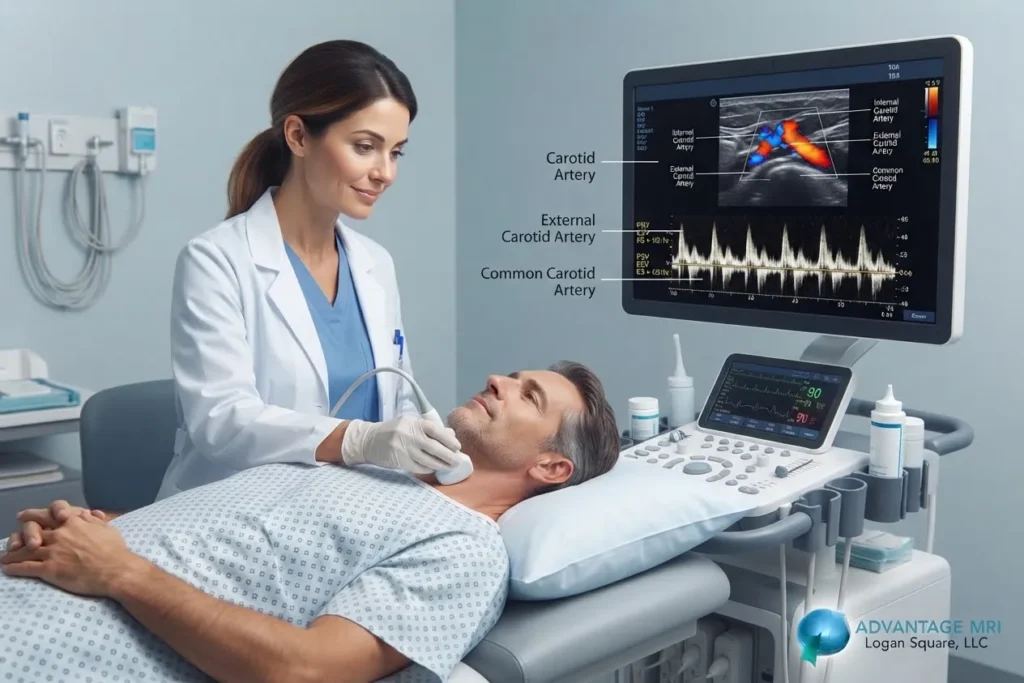
If your doctor just mentioned a carotid doppler ultrasound and you left the office with more questions than answers, you’re not alone. It’s one of those tests that sounds complicated but is actually pretty simple once someone walks you through it. And the truth is, it’s one of the best tools doctors have for catching artery problems before they turn into something serious like a stroke.
Here’s the thing. Your carotid arteries carry blood from your heart to your brain and when they start to narrow, you usually don’t feel a thing. That’s why this test matters so much. A carotid doppler ultrasound lets a radiologist look at blood flow through those arteries in real time, without needles, radiation or any real discomfort.
At Advantage MRI in the Hermosa neighborhood of Chicago, we run this exam regularly for patients whose doctors want a closer look at their vascular health. Below we’ll break down what the test checks, how it compares to a transcranial doppler ultrasound, what happens on the day of your appointment and how to make sense of your results once they come in.
What Is a Carotid Doppler Ultrasound
A carotid doppler ultrasound is a non invasive imaging test that uses sound waves to check blood flow through your carotid arteries. These are the two large blood vessels on each side of your neck that supply blood to your brain, face and neck.
The test works through something called the Doppler effect. A handheld device called a transducer sends sound waves into your neck and picks up how those waves bounce off moving blood cells. The machine translates that information into images and sound, which lets the technologist see how fast blood is moving and whether anything is blocking its path.
This kind of scan is often called a carotid duplex ultrasound because it combines two types of imaging at once. Regular ultrasound shows the structure of the artery, while Doppler ultrasound shows the blood flow moving through it. Together they give a much fuller picture than either one alone.
Plain and simple, this test is looking for narrowing, called carotid artery stenosis and buildup of plaque along the artery walls. Plaque is made up of fat, cholesterol and other substances that stick to the inside of blood vessels over time. When enough of it builds up, it can restrict blood flow or break loose and travel toward the brain.
| Artery | Location | What It Supplies |
|---|---|---|
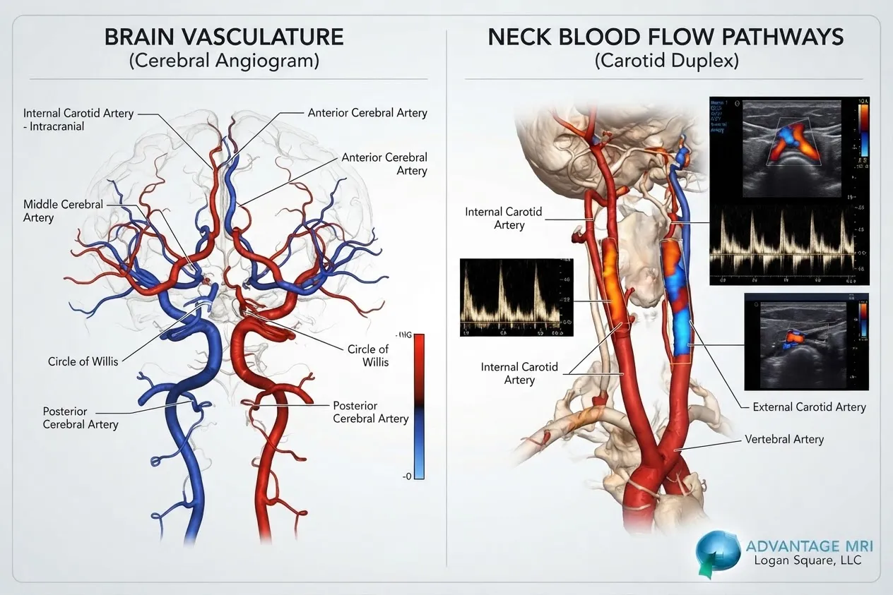
| Common carotid artery | Runs up each side of the neck | Splits into internal and external branches |
| Internal carotid artery | Branches off at the carotid bifurcation | Blood flow to the brain |
| External carotid artery | Branches off alongside the internal carotid | Blood flow to the face and scalp |
The internal carotid artery is usually the one doctors care about most, since narrowing there raises stroke risk more directly than narrowing in the external branch.
Want to know how this compares to imaging tests like MRI imaging? MRI gives a detailed picture of tissue and structure, while carotid doppler focuses specifically on blood flow and vessel narrowing. Doctors sometimes order both depending on what they’re trying to rule out.
Why Doctors Order Carotid Doppler Ultrasound
Doctors don’t order this test just because. There’s usually a reason and it almost always ties back to stroke prevention.
Carotid artery disease develops slowly. Plaque builds up over years, often without any symptoms at all. For a lot of people, the first sign something’s wrong is a mini stroke, also called a transient ischemic attack or a full stroke. That’s exactly what this test tries to catch before it happens.
A few common reasons your doctor might send you for this exam:
- Your doctor heard a whooshing sound over your neck artery during a checkup, known as a carotid bruit
- You’ve had symptoms like sudden weakness, numbness, vision changes or trouble speaking
- You have risk factors like high blood pressure, high cholesterol, diabetes or a history of smoking
- You’ve already been diagnosed with coronary artery disease or peripheral artery disease elsewhere in your body
- You have a family history of stroke or carotid artery disease
Heart and vascular health tend to travel together. If you’ve already had an EKG test or been checked with an echocardiogram for heart concerns, your doctor might follow up with a carotid ultrasound too, since artery disease in one part of the body often shows up somewhere else.
Carotid Doppler vs Transcranial Doppler Ultrasound
People mix these two up a lot and honestly, it makes sense. Both use Doppler technology and both look at blood flow to the brain. But they’re checking different parts of the pathway.
A carotid doppler ultrasound scans the arteries in your neck, outside the skull. A transcranial doppler ultrasound goes further, checking blood flow through arteries inside the skull itself, using a thinner sound wave that can pass through bone.
| Feature | Carotid Doppler Ultrasound | Transcranial Doppler Ultrasound |
|---|---|---|
| Area scanned | Neck arteries (extracranial carotid) | Arteries inside the skull |
| What it checks | Stenosis, plaque, blood flow velocity | Blood flow speed and direction in brain vessels |
| Common reason ordered | Stroke risk screening, carotid bruit | Sickle cell disease, vasospasm, stroke monitoring |
| Typical exam time | 30 to 45 minutes | 30 to 60 minutes |
In a lot of cases, doctors order both together when they want a complete look at blood flow from the neck all the way into the brain. This combined approach is sometimes referred to as carotid and transcranial doppler ultrasound imaging.
What Happens During a Carotid Doppler Ultrasound
Not much prep needed here and that’s part of why patients tend to like this test.
Before the test. No fasting is required. You can eat and drink normally beforehand. Wear something comfortable with a collar you can move out of the way, since the technologist needs clear access to both sides of your neck.
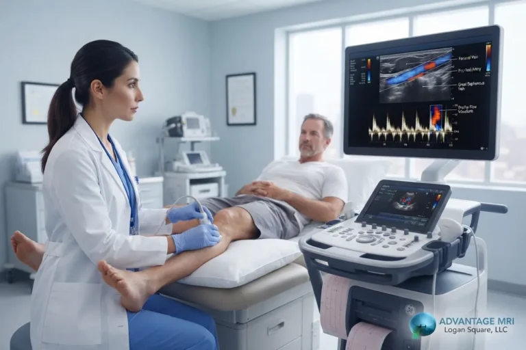
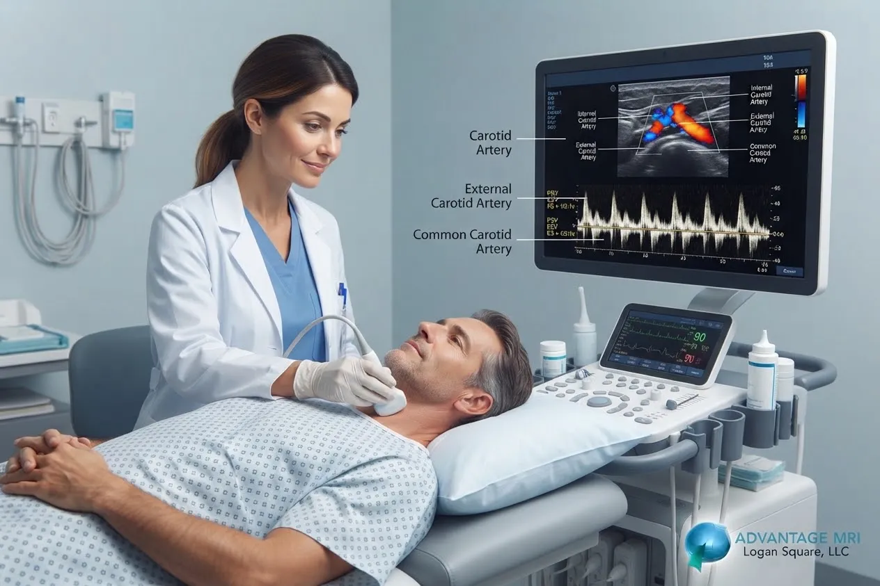
During the scan. You’ll lie down on the exam table with your head turned slightly to one side. The technologist applies a warm gel to your neck, which helps the sound waves transmit clearly. Then they move the transducer along your neck, pausing at certain points to capture images and record the sound of your blood flow. You might hear a rhythmic whooshing noise through the machine’s speaker. That’s completely normal and it’s just the sound of blood moving through your artery.
After the test. There’s no recovery time needed. You can drive yourself home and go right back to your normal day. The technologist wipes off the gel and you’re done.
A board certified radiologist reviews the images afterward and prepares a report for your referring doctor. At Advantage MRI, exams are reviewed by radiologists including Dr. Gregory T. Goldstein, who has years of experience in vascular and diagnostic imaging.
Is Carotid Doppler Ultrasound Painful or Risky
No. This is one of the safer, easier tests out there and it’s worth saying plainly.
There’s no needle involved and no radiation exposure, unlike a CT scan or X ray. The transducer just glides along your skin with a bit of gentle pressure. Most patients describe it as mildly uncomfortable at worst, mainly from having to hold your neck in one position for a few minutes.
Since there’s no radiation, this test is considered safe for repeat monitoring, which matters if your doctor wants to track how your carotid arteries change over time. The American College of Radiology recognizes ultrasound as one of the safest imaging tools available precisely because it doesn’t rely on radiation at all.
Understanding Your Carotid Doppler Ultrasound Results
This is usually where things get confusing, so let’s slow down here.
Your results will focus on something called peak systolic velocity, which measures how fast blood is moving through your artery at its fastest point during each heartbeat. Faster than normal flow through a narrow section usually points to a blockage, since blood speeds up when it’s forced through a tighter space, kind of like water through a narrowed hose.
Radiologists use velocity criteria developed by groups like the Society of Radiologists in Ultrasound Consensus Conference to grade how much narrowing is present.
| Stenosis Grade | Approximate Blockage | What It Generally Means |
|---|---|---|
| Mild | Less than 50% | Usually monitored, often no immediate treatment |
| Moderate | 50% to 69% | May need medication, lifestyle changes or closer monitoring |
| Severe | 70% or higher | May require further evaluation for procedures like carotid endarterectomy or carotid artery stenting |
These numbers are general guidelines, not a diagnosis on their own. Your doctor will look at your full health picture, including symptoms and other test results, before deciding on next steps. This article isn’t a substitute for medical advice and any results should be reviewed with your own physician.
Who Should Get a Carotid Artery Ultrasound
Not everyone needs this test and that’s actually an important point. Screening someone with no symptoms and no risk factors isn’t usually recommended, since it can lead to unnecessary follow up testing for findings that were never going to cause a problem.
That said, a lot of people fall into groups where this test genuinely makes sense:
- Adults over 65, especially with a history of smoking or high blood pressure
- People who’ve had a stroke or transient ischemic attack already
- Anyone with known coronary artery disease or peripheral artery disease
- Patients whose doctor detected a carotid bruit during a physical exam
- People with a strong family history of stroke at a younger age
Risk tends to climb with age but it’s not the only factor. Diabetes, smoking history and cholesterol levels all play a role too. If a few of these apply to you, it’s worth bringing up with your doctor rather than assuming you’re fine.
Carotid Doppler Ultrasound in Chicago at Advantage MRI
Advantage MRI has been serving the Hermosa neighborhood and greater Chicago area with accredited diagnostic imaging and carotid doppler ultrasound is one of the vascular exams we perform regularly.
Our center is accredited by the American College of Radiology, which means our equipment, staff qualifications and imaging protocols meet national standards for quality and safety. Every scan is reviewed by a board certified radiologist and reports go back to your referring physician quickly so there’s no unnecessary waiting around.
We also offer on site pharmacy access and IV treatment capability for patients who need it, along with fast scheduling for both routine and urgent referrals. If your doctor has ordered a carotid doppler ultrasound or if you’re wondering whether you should ask about one, our team at Advantage MRI can help walk you through the process from scheduling to results.
Related imaging services we offer include digital X ray, EMG testing and holter monitor exams for patients whose care involves more than one type of diagnostic workup.
Frequently Asked Questions
What does a carotid doppler ultrasound check for?
It checks blood flow through your carotid arteries and looks for narrowing or plaque buildup that could raise your risk of stroke. It also measures how fast blood moves through the artery, which helps radiologists spot blockages.
How long does a carotid doppler ultrasound take?
Most exams take between 30 and 45 minutes. Some cases take a bit longer if the technologist needs extra images from both sides of the neck.
Do I need to fast before a carotid doppler ultrasound?
No. You can eat and drink normally before this test. There’s no special prep needed beyond wearing comfortable clothing that allows access to your neck.
Is a carotid doppler ultrasound painful?
No. It’s a non invasive test with no needles and no radiation. Most people feel nothing more than mild pressure from the transducer moving across the skin.
What’s the difference between carotid doppler and doppler neck ultrasound?
They’re generally the same thing. Doppler neck ultrasound is just another way people refer to a carotid doppler exam, since the carotid arteries run through the neck.
How accurate is carotid doppler ultrasound for detecting blockage?
It’s considered a reliable first step for detecting carotid artery stenosis, especially when interpreted using standardized velocity criteria. In some cases, doctors may follow up with additional imaging like angiography for further confirmation.
What is considered a normal carotid doppler reading?
A normal reading shows smooth, steady blood flow with no significant narrowing and peak systolic velocity within expected ranges for a healthy artery. Your radiologist’s report will note whether your results fall within normal limits.
Can a carotid doppler ultrasound detect stroke risk before symptoms appear?
Yes, that’s actually the main reason this test exists. Since carotid artery disease often develops without symptoms, this scan can catch narrowing early, before it leads to a stroke or transient ischemic attack.
How often should high risk patients get carotid screening?
This depends entirely on your individual risk factors and prior results, so it’s a conversation to have with your doctor. Patients with known moderate to severe stenosis are often monitored more frequently than those with mild or no narrowing.
When will I get my carotid doppler ultrasound results?
A radiologist reviews your images and sends a report to your referring doctor, often within a day or two. Your doctor’s office will then discuss the findings with you directly.
Conclusion
A carotid doppler ultrasound is a quick, painless way to check on something that matters a lot more than most people realize, the blood flow keeping your brain supplied and healthy. It won’t fix a problem on its own but catching narrowing or plaque buildup early gives you and your doctor a real head start on managing it. If your doctor has recommended this test, there’s really no reason to put it off. And if you’re just curious whether it’s right for you, that’s a good conversation to bring to your next appointment.
This article is for general information only and isn’t a substitute for professional medical advice, diagnosis or treatment. Always talk to your doctor about your specific health needs. For reliable, advanced diagnostic imaging you can trust, call Advantage MRI today or schedule your appointment online.
Contact Us
- Address: 2008 N Pulaski Rd, Chicago, IL 60639
- Phone: (773) 227 9777
- Website: Contact Advantage MRI
Follow us on social media for the latest health tips, diagnostic imaging updates and clinic news: Facebook | Instagram | LinkedIn.