Health & Imaging
Ultrasound with Doppler Vascular: What the Test Shows and What to Expect

Your doctor said the words doppler ultrasound and now you are sitting there wondering what that actually means for your body. Maybe your legs have been swelling. Maybe there is a family history of blood clots and someone wants to rule that out. Whatever brought you here the good news is this test is painless and it does not involve any needles or radiation.
An ultrasound with doppler vascular study is a way for doctors to see how blood is moving through your veins and arteries. It uses sound waves instead of dye or radiation which makes it one of the safer imaging options available today. Below you will find a plain English breakdown of what the test involves how it works what it can catch and what it costs.
Quick answer: An ultrasound with doppler vascular exam checks blood flow direction and speed inside your veins and arteries using sound waves. It takes 20 to 45 minutes, involves no needles, and helps doctors spot clots, narrowed arteries, and valve problems.
What Is a Vascular Doppler Ultrasound
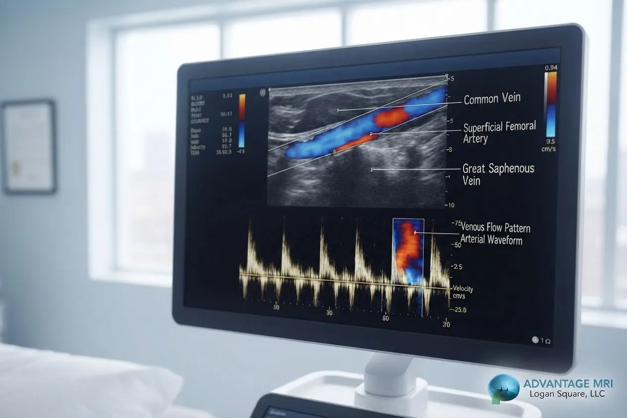
A vascular doppler ultrasound is a non-invasive test that uses high-frequency sound waves to look at blood flow through veins and arteries. A handheld device called a transducer sends sound waves into the body. Those waves bounce off moving red blood cells and come back changed based on how fast and in what direction the blood is traveling. That change is called the doppler shift and it is what lets a sonographer see blood flow in real time.
Unlike an x-ray or CT scan there is no radiation involved. The test is also completely painless. Most people describe it as feeling like a regular ultrasound with a bit of pressure from the transducer moving across the skin.
Doctors order this test to check for blood clots poor circulation narrowed arteries and a handful of other vascular concerns. It is one of the more common tools used in vascular medicine because it gives a clear picture without putting the patient through anything invasive.
How a Doppler Ultrasound Differs From a Regular Ultrasound
Here is the thing people often mix up. A regular ultrasound and a doppler ultrasound are not entirely separate tests. They actually work together most of the time.
A standard ultrasound shows the structure of an organ or tissue. Think shape size and any visible abnormality. Doppler ultrasound adds a layer on top of that by measuring how blood is moving through vessels. When both are combined the exam is often called a duplex ultrasound because it does double duty showing structure and flow at the same time.
| Feature | Standard Ultrasound | Doppler Ultrasound |
|---|---|---|
| What it shows | Shape and structure of organs or tissue | Direction and speed of blood flow |
| Common use | Checking organs, soft tissue, pregnancy | Checking veins, arteries, circulation |
| Sound technology | Basic sound wave reflection | Measures doppler shift in moving blood cells |
| Often paired with | Doppler for full vascular picture | Standard imaging for duplex scanning |
So when someone says they are getting a vascular ultrasound test they are usually getting both types rolled into one appointment.
What a Vascular Doppler Ultrasound Can Detect
This test covers a fair amount of ground. Doctors use it to look for several different vascular problems and honestly that is part of why it gets ordered so often.
Blood clots and deep vein thrombosis. A clot in a deep vein, especially in the leg, can be dangerous if it breaks loose. Doppler ultrasound is one of the main tools doctors use to spot a DVT early.
Artery blockages and plaque buildup. Fatty deposits called plaque can narrow an artery over time. This narrowing reduces blood flow and doppler imaging can show exactly where that reduction is happening.
Varicose veins and poorly functioning valves. When vein valves stop working correctly, blood can pool instead of flowing back toward the heart. This is known as venous insufficiency and doppler ultrasound is a standard way to confirm it.
Aneurysms. An aneurysm is a bulge in a weakened artery wall. Doppler imaging can help identify the size and location so a doctor can decide on next steps.
Vascular malformations. These are abnormal connections between arteries and veins, sometimes called arteriovenous malformations, and doppler flow studies can help map them out.
Types of Vascular Doppler Studies
Not every doppler ultrasound looks at the same part of the body. The type of study depends on what your doctor is trying to rule out.
| Study Type | Area Checked | Typical Reason for Testing |
|---|---|---|
| Venous Doppler | Legs, arms | Blood clots, venous insufficiency, swelling |
| Arterial Doppler | Legs, arms | Peripheral arterial disease, poor circulation |
| Carotid Doppler | Neck arteries | Stroke risk, plaque buildup |
Extremity studies are the most common, meaning the lower extremities or upper extremities depending on where the symptoms are showing up. A carotid doppler focuses on the arteries in the neck that supply blood to the brain, which is a different concern entirely from leg pain or swelling.
Some vascular labs also use something called a PVR, or pulse volume recording, alongside doppler imaging when they need segmental pressure readings along the leg. This is more common in cases where peripheral arterial disease is already suspected.
Vascular Doppler Ultrasound vs Other Vascular Tests
People sometimes ask whether an ultrasound with doppler vascular exam is really the best first step compared to other options like a CT angiogram or MRI. Here is a simple way to think about it.
- Doppler ultrasound is noninvasive, has no radiation, and is usually the first test ordered because it is quick and low cost compared to advanced imaging.
- CT angiogram uses radiation and contrast dye and gives a more detailed structural view, often used when doppler results are unclear or a more precise map is needed before a procedure.
- MRI imaging avoids radiation like ultrasound but takes longer and costs more, and it is typically reserved for cases needing very detailed soft tissue or vessel imaging.
Most vascular specialists start with doppler ultrasound because it answers the basic question of how blood is flowing without exposing the patient to anything invasive. If the findings are unclear or more detail is needed, a doctor may then order additional imaging or refer the patient for more advanced vascular studies.
What Happens During the Test
Walking into your appointment not knowing what to expect can make anyone nervous. Here is what actually happens step by step.
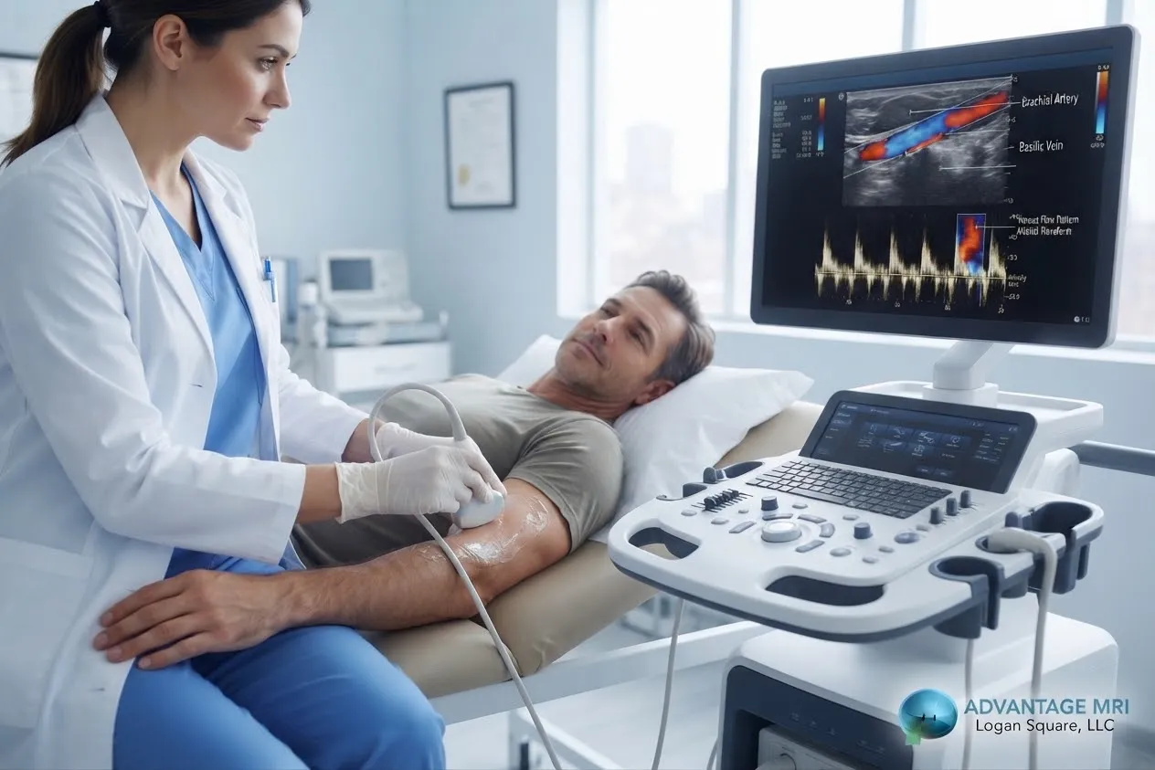
- Check in and get comfortable. You will be asked to lie down and the area being examined, whether it is your leg, arm, or neck, will be exposed.
- Gel goes on the skin. A sonographer applies a small amount of gel to help the transducer glide smoothly and pick up a clear signal.
- The transducer does the work. The sonographer moves the handheld transducer over the area, pausing at different points to capture images and flow readings. You might hear a whooshing sound during this part, which is just the sound of blood flow being picked up by the machine.
- Wrapping up. The gel gets wiped off and that is it. There is no recovery time needed.
The whole appointment usually takes somewhere between 20 and 45 minutes depending on how many areas are being checked. A brachial pressure reading might also be taken in the arm for comparison if arterial flow is being evaluated.
One thing worth mentioning is that the portability of ultrasound equipment means this test can sometimes be done bedside for patients who cannot easily move, though most people will have it done in a dedicated imaging room.
How to Prepare for a Venous Doppler Ultrasound
Preparation for this test is fairly simple compared to other imaging exams.
Wear loose comfortable clothing since you may need to expose your legs or arms. Avoid lotion or oil on the skin the day of the test since it can interfere with the gel and transducer contact. If you are a smoker you may be asked to avoid nicotine for a couple hours before an arterial study since it can temporarily affect blood flow readings.
There is generally no fasting required and most people can take their usual medications. That said, always follow the specific instructions given by your doctor or the imaging center since individual cases can vary.
Understanding Your Doppler Ultrasound Results
A normal doppler ultrasound will show blood moving smoothly in the expected direction without narrowing, blockage, or pooling. The sonographer captures the images and flow data, but it is a radiologist who reviews everything and writes the formal report.
If something abnormal shows up, like a clot, reduced flow, or a valve that is not working right, the report will note where it was found and how severe it looks. Your doctor will then use that information to figure out next steps, which could mean medication, further imaging, or a referral to a vascular specialist.
At Advantage MRI, results are reviewed by board-certified radiologists, including founder Dr. Gregory T. Goldstein, MD, who specializes in diagnostic imaging interpretation. Having an experienced radiologist read the waveform analysis and flow data matters because subtle findings can be easy to miss without the right training.
This information is meant to help you understand the process and is not a substitute for medical advice from your own physician.
Doppler Ultrasound Cost
Doppler ultrasound price can vary quite a bit depending on a few factors, so there is no single number that applies to everyone.
What affects the price:
- Which body area is being scanned and how many areas are included
- Whether insurance is being used or the visit is self-pay
- Whether the study includes additional testing like a PVR alongside the doppler exam
Insurance and self-pay options. Most insurance plans cover vascular doppler ultrasound when it is ordered by a doctor for a documented medical reason. For patients without insurance or with high deductibles, self-pay pricing is usually available and it helps to ask the imaging center directly for a quote based on your specific study.
The best way to get an accurate number is to call ahead and ask about your specific situation since insurance coverage and self-pay rates differ from person to person.
It also helps to ask whether the quoted price covers the full study or just the scan itself. Some centers bill the technical portion, meaning the equipment and sonographer time, separately from the professional fee, which covers the radiologist reading and reporting the results. Asking this upfront avoids surprise billing later.
Who Should Consider a Vascular Ultrasound
Doctors typically order this test when a patient has certain symptoms or risk factors that point toward a circulation problem.
Common symptoms that lead to testing include leg swelling, pain or cramping while walking, visible varicose veins, skin discoloration around the ankles, or one leg feeling warmer and more tender than the other.
Risk factors worth mentioning to your doctor include a family history of blood clots, recent surgery or long periods of immobility, smoking, diabetes, and a personal history of vascular disease.
If any of this sounds familiar, bringing it up with your doctor is the first step. They will decide whether an ultrasound with doppler vascular exam makes sense for your situation.
It is worth adding that not every leg pain or swelling case points to a vascular problem. Sometimes symptoms overlap with nerve issues or muscle strain, which is part of why doctors use a combination of history, physical exam, and imaging before landing on a diagnosis.
Getting a Vascular Doppler Ultrasound in Chicago
Advantage MRI is located in the Hermosa neighborhood of Chicago and offers vascular ultrasound imaging along with a full range of diagnostic services. The center is ACR accredited, which means it meets national standards for equipment, safety, and image quality set by the American College of Radiology.
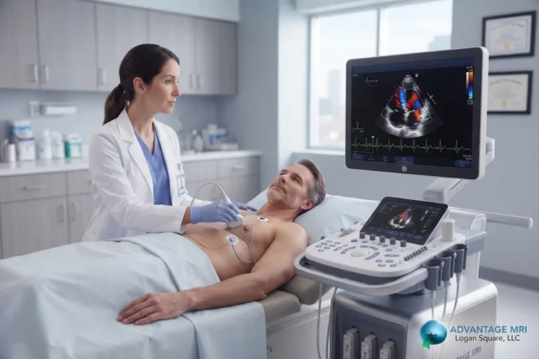
Since circulation issues can sometimes overlap with heart related symptoms, doctors may also request an echocardiogram or EKG testing to rule out cardiac causes. For patients being monitored over a longer period, a holter monitor may be used alongside vascular imaging to get a fuller picture of heart rhythm and circulation together.
Leg pain and numbness are not always vascular either. Sometimes a doctor wants to rule out nerve involvement with an EMG test or a nerve conduction study before settling on a vascular diagnosis. And in cases where a deeper structural look is needed, MRI imaging can provide additional detail beyond what ultrasound alone can show.
Advantage MRI works closely with referring physicians and offers same day scheduling in many cases along with fast reporting turnaround. You can read more about the radiology team on the about us page.
Frequently Asked Questions
What is a vascular doppler ultrasound used for?
It is used to check blood flow through veins and arteries, helping doctors detect blood clots, blocked or narrowed arteries, and valve problems that affect circulation.
Is a doppler ultrasound painful?
No. It is a non-invasive test that involves gel and a handheld transducer moving across the skin. Most patients feel nothing more than light pressure.
How long does a vascular doppler ultrasound take?
Most exams take between 20 and 45 minutes depending on how many areas of the body are being examined.
Can a doppler ultrasound find a blood clot?
Yes. Venous doppler studies are one of the primary tools doctors use to detect deep vein thrombosis in the legs and arms.
What is the difference between venous and arterial doppler?
A venous doppler checks blood flow in the veins, often looking for clots or valve problems. An arterial doppler checks blood flow in the arteries, often looking for narrowing or blockages tied to peripheral arterial disease.
Do I need to prepare anything before a venous doppler ultrasound?
Preparation is minimal. Wear loose clothing, skip lotion on the area being scanned, and follow any specific instructions given by your doctor or the imaging center.
How much does a doppler ultrasound cost?
Doppler ultrasound price depends on the body area being scanned, whether insurance is used, and whether additional testing is included. Calling ahead for a quote is the most accurate way to know your cost.
Who interprets doppler ultrasound results?
A board-certified radiologist reviews the images and flow data captured by the sonographer and writes the official report that goes to your doctor.
Is a doppler ultrasound the same as a CT scan?
No. A doppler ultrasound uses sound waves and involves no radiation, while a CT scan uses x-ray technology. Doctors often choose ultrasound first for vascular concerns because it is noninvasive and does not expose the patient to radiation.
When should I ask my doctor about a vascular ultrasound?
If you are dealing with leg swelling, pain while walking, visible varicose veins, or have risk factors like a family history of blood clots, it is worth bringing up with your doctor to see if testing makes sense.
Conclusion
A vascular doppler ultrasound gives doctors a clear noninvasive way to check how well blood is moving through your veins and arteries. Whether the concern is a possible clot, narrowed artery, or valve problem, this test offers useful answers without needles, radiation, or downtime. If you have symptoms like swelling, leg pain, or a family history of circulation problems, talking to your doctor about whether this test fits your situation is a reasonable next step.
This article is for general information and is not a substitute for professional medical advice, diagnosis, or treatment. Always talk to your doctor about your specific health concerns.
For reliable, advanced diagnostic imaging you can trust, call Advantage MRI today or schedule your appointment online.
Contact Us
- Address: 2008 N Pulaski Rd, Chicago, IL 60639
- Phone: (773) 227-9777
- Contact us online
Follow us on social media for the latest health tips, diagnostic imaging updates, and clinic news: Facebook | Instagram | LinkedIn.