Health & Imaging
How Is an Echocardiogram Done on a Woman? A Step-by-Step Walkthrough
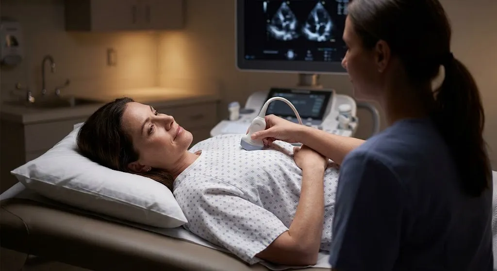
How is an echocardiogram done on a woman? A standard echocardiogram (transthoracic echo, or TTE) is done the same way for women as for men. You undress from the waist up, including your bra, and put on a gown that opens in the front. You lie mostly on your left side while a trained sonographer moves a gel-covered ultrasound probe over several spots on your chest: beside the breastbone, under the left breast near the tip of the heart, below the ribs and above the collarbone. To get clear images, the sonographer may need to gently move or ask you to lift your left breast. You stay covered except for the small area being scanned, and you can ask for a chaperone. The test is painless, uses no radiation, and usually takes 30 to 60 minutes.
Many women feel uneasy about the undressing and close contact involved. Knowing exactly what happens and what you can ask for makes the experience much easier. This guide walks through each step honestly and calmly.
What Is an Echocardiogram?
An echocardiogram is an ultrasound of the heart. It uses sound waves to create moving pictures of your heart’s chambers, walls and valves, and Doppler ultrasound to measure blood flow. It helps doctors see:
Need to Schedule a Diagnostic Appointment?
Whether you need an MRI, ultrasound, X-ray, or another diagnostic service, Advantage MRI is here to help you take the next step in your healthcare journey.
Submit your appointment request online, and our team will contact you to confirm your visit.
Book Your Appointment →- How well your heart pumps (often reported as the ejection fraction)
- The size of the heart chambers and the thickness of the walls
- How the four valves open and close, and whether they leak or are narrowed
- Fluid around the heart
- Clues to pressure in the lungs
For a complete overview of types, uses and results, see our pillar guide, What Is an Echocardiogram?
Why Is the Echocardiogram Procedure Different for Women?
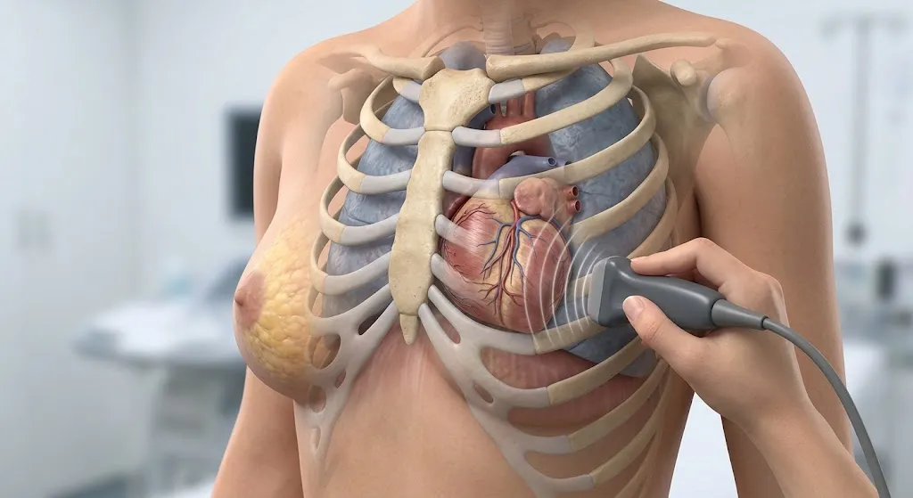
The test itself is identical. The difference is practical: the heart sits behind the left side of the chest, and in women, breast tissue covers part of the area where the best “windows” for imaging are. Ultrasound waves travel poorly through thick tissue, so the sonographer needs to place the probe directly on the chest wall, sometimes under or beside the left breast. That’s the main reason a woman’s echo involves more repositioning, and it’s a normal, professional part of the exam.
Before Your Appointment: How to Prepare
A standard transthoracic echocardiogram needs very little preparation:
- Eat and drink normally unless your doctor says otherwise. Fasting isn’t usually needed for a standard echo. (Stress echoes and transesophageal echoes have different instructions.)
- Take your usual medications.
- Wear a two-piece outfit, such as a top with pants or a skirt, so you only need to remove your top.
- Choose an easy-to-remove bra. You’ll take it off for the test.
- Skip lotion, oil or powder on your chest that day so electrodes and the probe make good contact.
- Leave necklaces at home or plan to remove them.
- Bring your ID, insurance card, doctor’s order and a medication list.
- Ask for accommodations when you book, such as a chaperone or, if available, a female sonographer.
The Echocardiogram Procedure, Step by Step
Step 1: Changing Into a Gown
You’ll be shown to a private room or changing area. You’ll remove clothing and your bra from the waist up and put on a gown with the opening in the front. You can keep your pants or skirt on. The sonographer will step out or turn away while you change.
Step 2: EKG Electrodes
Three small sticky electrodes are usually placed on your chest or shoulders. These connect to the ultrasound machine so it can time the images with your heartbeat. This is a simple monitoring strip, not a full EKG test. Learn how the two tests differ in our echocardiogram vs EKG guide.
Step 3: Positioning on Your Left Side
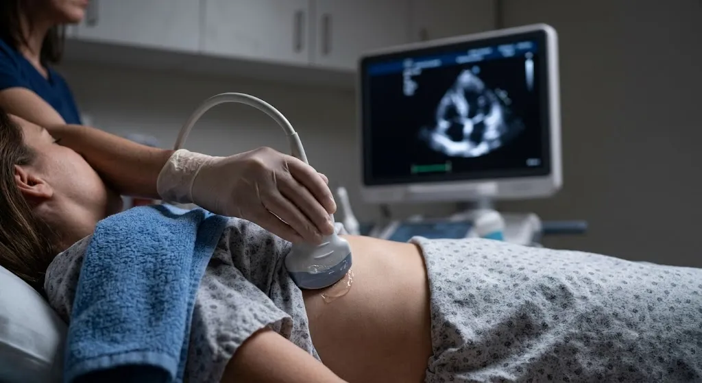
You’ll lie on an exam table, usually turned onto your left side, with your left arm raised and bent so your hand rests near or under your head. This position brings the heart closer to the chest wall and moves the lung out of the way, which gives clearer pictures. Some exam tables have a cutout so the probe can reach the chest from underneath.
The room is typically dimmed so the sonographer can see the screen clearly.
Step 4: Draping for Privacy
The sonographer should keep you covered with the gown and, often, a sheet or towel. Only the small area being scanned is exposed at any time. If you feel too exposed, say so. Adjusting the drape is easy.
Step 5: Gel and the Probe
Warm (or sometimes cool) gel is applied to the probe. The gel removes air between the probe and your skin, which ultrasound needs to work. The sonographer presses the probe against your chest. Firm pressure is normal because it improves the images.
Step 6: Imaging From Several Positions
The sonographer collects images from a series of standard “windows”:
| Window | Where the probe is placed | What it shows |
|---|---|---|
| Parasternal | Left of the breastbone, upper chest | Long and short views of the heart and valves |
| Apical | Near the tip of the heart, usually under or beside the left breast | All four chambers and valve flow |
| Subcostal | Just below the ribs, under the breastbone | Heart from below; fluid around the heart; the large vein to the heart |
| Suprasternal | Above the breastbone at the base of the neck | The aorta (main artery) |
Step 7: The Apical View and Breast Tissue
This is the step most women ask about. To capture the apical view, the probe needs to sit on the chest wall at the lower left side of the heart, which is often beneath the left breast. The sonographer will usually:
- Explain what they’re about to do
- Ask you to lift your left breast with your hand, or
- Gently move breast tissue aside with their hand or the probe
This is done to get a clear, accurate image, and it lasts only as long as needed. You’re always allowed to ask questions, ask for a pause, or ask for the step to be explained before it happens.
Step 8: Breathing Instructions and Position Changes
You may be asked to:
- Breathe out and hold your breath for a few seconds
- Take a slow breath in
- Roll onto your back for the subcostal and suprasternal views
- Tilt your head back slightly for the view above the breastbone
Step 9: Doppler Sounds
During Doppler imaging, the machine may make whooshing or pulsing sounds. That’s the sound of blood moving through your heart and valves. It’s completely normal.
Step 10: Contrast (Sometimes)
If images are hard to see, the sonographer may recommend an ultrasound enhancing agent given through an IV. These are tiny gas-filled microspheres that make the heart’s inner walls easier to see. They’re different from the contrast used in CT or MRI. Not every echo needs one.
Step 11: Finishing Up
The gel is wiped off, electrodes are removed, and you can get dressed. There’s no recovery time. You can drive, eat and return to your usual activities right away.
Does an Echocardiogram Hurt?
A standard echocardiogram doesn’t hurt. You may feel:
- Coolness or slipperiness from the gel
- Firm pressure from the probe, especially below the ribs and near the tip of the heart
- Mild discomfort if you have chest wall tenderness, recent surgery or very sensitive breasts
If the pressure is uncomfortable, tell the sonographer. They can adjust their technique.
Privacy Rights and Requests You Can Make
You’re entitled to dignity and comfort during any medical exam. Reasonable requests include:
- A chaperone in the room during the test
- A female sonographer, if one is available (ask when scheduling; it can’t always be guaranteed)
- Draping so only the scanned area is uncovered
- An explanation before each step that involves touching or repositioning
- A pause if you need a moment
- Keeping a support person with you, depending on facility policy
If something makes you uncomfortable, say so. A professional sonographer will respond respectfully.
Special Situations for Women
Breast Implants
Implants can block ultrasound waves and reduce image quality. Let the sonographer know beforehand so they can adjust angles and windows. Contrast may sometimes be recommended to improve images.
After Breast Surgery or Mastectomy
Scar tissue, radiation changes or reconstruction can affect imaging windows. Let the team know about past surgery, tenderness or areas to avoid.
Pregnancy
Echocardiograms are safe during pregnancy because ultrasound doesn’t use radiation. Doctors sometimes order echos in pregnancy to evaluate heart murmurs, shortness of breath or known heart conditions. Lying on your left side is also comfortable in later pregnancy. Ultrasound enhancing agents are generally used in pregnancy only when clearly needed.
Larger Body Size
More tissue between the probe and the heart can make imaging harder. The sonographer may use more pressure, different positions or contrast. This doesn’t mean anything is wrong. It’s just a technical challenge.
Chest Discomfort Lying on Your Side
If lying on your left side is difficult because of pain or a medical condition, tell the sonographer. They’ll find positions that work for you.
Why Women Are Commonly Referred for an Echocardiogram
Doctors order echocardiograms for many reasons. Some come up especially often for women:
- Palpitations or a racing heart, to check heart structure when rhythm symptoms are present
- Shortness of breath or fatigue, which can be early signs of heart disease in women
- A heart murmur heard during a routine exam, sometimes from a leaky valve or, in some cases, mitral valve prolapse
- Pregnancy-related concerns, such as breathlessness, swelling or high blood pressure in pregnancy, or symptoms in the months after delivery, when a rare condition called peripartum cardiomyopathy can occur
- Monitoring during certain cancer treatments, since some chemotherapy and targeted therapies (including some used for breast cancer) can affect heart function, and echocardiograms are often used to track it
- High blood pressure, to look for thickening of the heart muscle
- Chest pain, sometimes as part of a stress echocardiogram
- Autoimmune conditions such as lupus, which can affect the lining around the heart
Heart disease is a leading cause of death for women in the United States, according to the CDC, and symptoms in women can be less “classic” than in men. An echocardiogram is one of the most useful, non-invasive ways to look at the heart’s structure and function.
Managing Anxiety on Test Day
Feeling nervous is common, especially when undressing is involved or when you’re waiting on answers about your heart. A few strategies can help:
- Ask the sonographer to walk you through each step before it happens.
- Bring a support person if the facility allows it.
- Focus on slow breathing while lying on your side. It also helps the images.
- Remember that silence isn’t a bad sign. Sonographers concentrate on getting measurements and usually can’t comment on findings during the exam.
- Plan something calming afterward, and write down questions for your doctor.
How Long Does the Echocardiogram Procedure Take?
A standard transthoracic echocardiogram usually takes 30 to 60 minutes. Factors that can lengthen it include difficult imaging windows, the need for contrast, or a more detailed exam ordered by your doctor. Our article on how long an echocardiogram takes covers the full appointment timeline.
Other Types of Echocardiograms
Most people have a transthoracic echo as described above. Your doctor may order a different type:
- Transesophageal echocardiogram (TEE): A thin probe is passed down the throat under sedation to view the heart from behind. It’s used for detailed views of valves and clots. Fasting is required. See our TTE vs TEE comparison.
- Stress echocardiogram: Images are taken before and after exercise or medication that makes the heart work harder. See our guide to stress echocardiograms vs stress EKGs.
Getting Your Results
The sonographer records images and measurements but usually can’t give results during the exam. A physician interprets the study and sends a report to your referring doctor, who will discuss it with you. Results often include ejection fraction, chamber sizes, wall thickness, valve function and estimated lung pressures. For questions about what an echo can and can’t detect, read can an echocardiogram detect a blocked artery?
What Happens After Your Results
If your echo is normal, your doctor may focus on other explanations for your symptoms or simply reassure you. If it shows a finding such as a leaky valve, reduced pumping or thickened walls, the next steps might include medication changes, a repeat echo in six to twelve months to track changes, a stress test, rhythm monitoring or a cardiology referral. Ask your doctor whether your result needs follow-up and when. Keep a copy of the report, since future echos are most useful when they can be compared with earlier ones.
Echocardiograms at Advantage MRI in Chicago
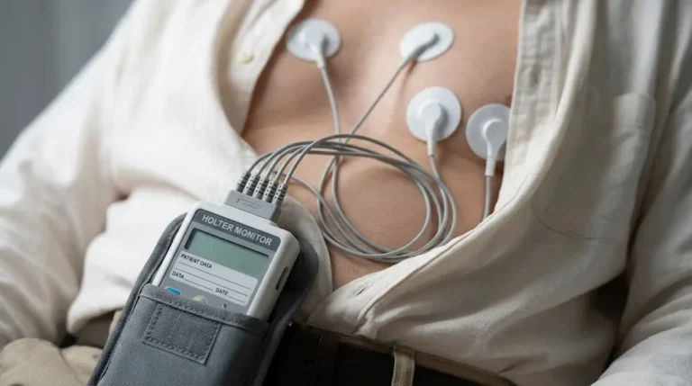
Advantage MRI performs echocardiograms at 2008 N Pulaski Rd, Chicago, IL 60639, along with EKGs, Holter monitoring, ultrasound, MRI, X-ray, EMG and nerve conduction studies. Our website emphasizes a comfortable, stress-conscious patient experience. If you’d like a chaperone or have other privacy preferences, tell us when you schedule so we can do our best to accommodate you.
We work with referring physicians and accept insured, out-of-network and self-pay patients.
Frequently Asked Questions About How Is An Echocardiogram Done On A Woman
Do I have to take my bra off for an echocardiogram?
Yes, in most cases. The probe needs direct contact with your chest, including under the left breast, and a bra gets in the way. You’ll wear a front-opening gown and stay draped.
Why do they touch your breast during an echocardiogram?
To get the apical view of the heart, the probe must sit on the chest wall at the lower left of the heart, often beneath the left breast. The sonographer may ask you to lift your breast or gently move it to get clear images.
Can I ask for a female sonographer?
You can ask. Availability depends on staffing, so request it when you book. You can also request a chaperone.
Can I eat before an echocardiogram?
For a standard transthoracic echo, usually yes. Fasting is typically needed only for a transesophageal echo or certain stress tests.
Is an echocardiogram safe during pregnancy?
Yes. It uses ultrasound, not radiation, and is commonly performed during pregnancy when needed.
Do breast implants affect an echocardiogram?
They can reduce image quality. Tell the sonographer beforehand so they can adjust positions or consider contrast.
Will the sonographer tell me my results?
Usually not. A physician interprets the images and sends the report to your doctor, who will review the results with you.
Conclusion
An echocardiogram for women follows the same steps as for anyone else, but it often involves more repositioning around the left breast to reach the best imaging windows. You’ll change into a front-opening gown, lie on your left side, and stay draped while a sonographer images your heart from several angles. The test is painless, radiation-free and usually done in under an hour. Knowing what to expect, and knowing you can ask for a chaperone, explanations or pauses, can make the appointment much more comfortable.
Need an echocardiogram in Chicago? Book an appointment with Advantage MRI or call (773) 227-9777. Hours: Monday-Friday 9:00 AM-5:00 PM, Saturday 9:00 AM-1:00 PM. Follow us on Facebook and Instagram.