Health & Imaging
What Is an EKG Test? How It Works and What It Detects
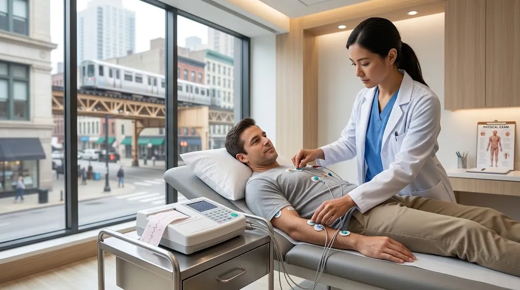
What is an EKG test? Quick answer: An EKG (electrocardiogram, also called an ECG) is a quick, painless test that records the electrical activity of your heart. Small sticky electrodes on your chest, arms and legs detect the tiny electrical signals that trigger each heartbeat, and a machine displays them as a wave pattern. A standard resting EKG records for about 10 seconds and takes 5 to 10 minutes in total. It helps doctors check your heart rate and rhythm and look for signs of arrhythmias, heart attacks (current or past), reduced blood flow, enlarged heart chambers, conduction problems, electrolyte imbalances and medication effects. It uses no radiation and doesn’t send electricity into your body.
This pillar guide covers what an EKG is, how it works, what it can and can’t detect, the different types of EKG tests, how to prepare, what results mean, and what happens next.
Need to Schedule a Diagnostic Appointment?
Whether you need an MRI, ultrasound, X-ray, or another diagnostic service, Advantage MRI is here to help you take the next step in your healthcare journey.
Submit your appointment request online, and our team will contact you to confirm your visit.
Book Your Appointment →EKG vs ECG: Is There a Difference?
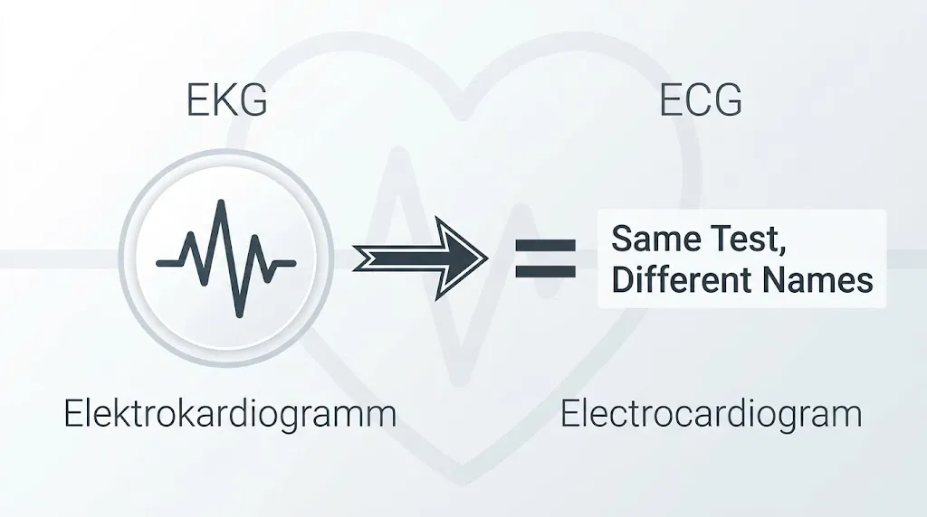
No. EKG and ECG are the same test. ECG comes from the English “electrocardiogram.” EKG comes from the German spelling, “Elektrokardiogramm,” and is widely used in the United States, partly to avoid confusion with EEG, a brain-wave test.
How the Heart’s Electrical System Works
To understand an EKG, it helps to know how your heartbeat starts:
- The sinoatrial (SA) node, a cluster of cells in the right upper chamber (atrium), acts as the heart’s natural pacemaker. It fires an electrical signal.
- The signal spreads across both atria, causing them to contract and push blood into the lower chambers.
- The signal reaches the atrioventricular (AV) node, which briefly delays it so the ventricles can fill.
- It then travels down the bundle of His and the right and left bundle branches.
- Finally, it spreads through Purkinje fibers into the ventricles, which contract and pump blood to the lungs and body.
Each of these steps shows up as part of the EKG wave pattern.
Reading the EKG Waves: P, QRS and T
| Part of the EKG | What it represents |
|---|---|
| P wave | Electrical activation of the atria (upper chambers) |
| PR interval | Time from atrial activation to the start of ventricular activation, including the AV node delay |
| QRS complex | Electrical activation of the ventricles (lower chambers) |
| ST segment | The period between ventricular activation and recovery; changes can suggest reduced blood flow or injury |
| T wave | Recovery (repolarization) of the ventricles |
| QT interval | Total time for ventricular activation and recovery |
A physician reviews these features across 12 views (leads) to build a picture of how your heart’s electrical system is working.
What Is a 12-Lead EKG?
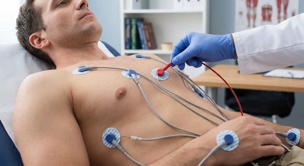
The standard EKG is called a 12-lead EKG, but it uses 10 electrodes:
- 4 on the limbs (one on each arm and leg)
- 6 across the chest
The machine combines signals from these electrodes to create 12 different “leads,” or viewpoints. Different leads look at different walls of the heart, which helps doctors locate problems such as which area of the heart may be affected by reduced blood flow.
What Does an EKG Test For? What It Can Detect
An EKG can help detect or provide clues about:
Heart Rhythm Problems (Arrhythmias)
- Atrial fibrillation and atrial flutter
- Premature beats (PACs and PVCs)
- Supraventricular tachycardia (SVT)
- Ventricular tachycardia
- Slow heart rhythms (bradycardia)
Heart Attack and Reduced Blood Flow
- Signs of a heart attack happening now, which are treated as an emergency
- Signs of a past heart attack, sometimes one that went unnoticed
- ST-segment and T-wave changes that may suggest reduced blood flow (ischemia)
Conduction Problems
- Heart block (first-, second- or third-degree AV block)
- Right or left bundle branch block
- Pre-excitation patterns such as Wolff-Parkinson-White
Structural Clues
- Enlargement or thickening of heart chambers (suggested, not measured directly)
- Inflammation of the lining around the heart (pericarditis)
Other Conditions and Effects
- Electrolyte imbalances, such as high or low potassium
- Effects of medications, such as QT prolongation
- Pacemaker function
- Some inherited electrical conditions
What an EKG Can’t Detect
An EKG is a powerful screening tool, but it has limits:
- It captures only about 10 seconds. Rhythm problems that come and go may be missed. That’s where Holter or event monitors help.
- It doesn’t show blocked arteries directly. A resting EKG is often normal in people with stable coronary artery disease. Stress testing or other imaging may be needed.
- It doesn’t show heart structure in detail. Valve problems, pumping strength and exact chamber size require an echocardiogram.
- It can’t predict every future heart attack.
Our guide to echocardiogram vs EKG explains how these two tests work together.
Why Would a Doctor Order an EKG?
Common reasons include:
- Chest pain or pressure
- Palpitations, a racing or irregular heartbeat
- Shortness of breath
- Dizziness, lightheadedness or fainting
- Unexplained fatigue
- High blood pressure, diabetes or high cholesterol
- Family history of heart disease or sudden cardiac death
- Before surgery (pre-operative assessment)
- Starting or monitoring certain medications
- Monitoring a known heart condition or pacemaker
- Routine baseline in some physical exams
If you have chest pain, fainting, severe shortness of breath or symptoms of a heart attack or stroke, call 911. Emergency teams perform EKGs immediately.
Types of EKG Tests
The term “EKG” covers several related tests:
| Type | How it works | Duration | Best for |
|---|---|---|---|
| Resting 12-lead EKG | Recorded lying down at rest | About 10 seconds of recording | Snapshot of rhythm, conduction and heart attack signs |
| Exercise stress EKG | Recorded while walking on a treadmill | Often about an hour for the full visit | Checking for reduced blood flow during exertion |
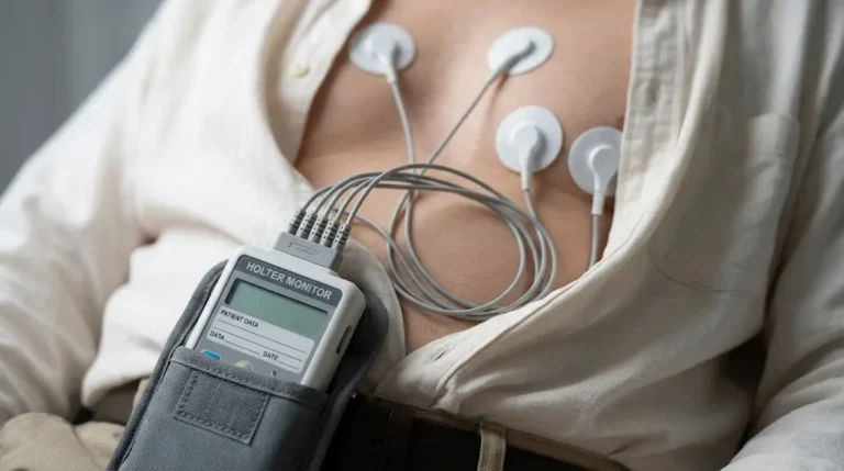
| Holter monitor | Portable recorder worn at home | 24-48 hours, sometimes up to 14 days | Frequent symptoms, counting irregular beats |
| Event monitor | Records when symptoms occur, or auto-detects | Weeks | Infrequent symptoms |
| Patch monitor | Adhesive patch records continuously | Often 1-2 weeks | Extended rhythm monitoring |
| Implantable loop recorder | Small device placed under the skin | Up to years | Rare but significant symptoms |
To learn more:
- Stress echocardiograms vs stress EKGs
- What is a Holter monitor?
- Holter monitor vs event monitor vs Zio patch
How to Prepare for an EKG
A resting EKG needs very little preparation:
- Avoid lotion, oil or powder on your chest, arms and legs.
- Wear a two-piece outfit so you only need to remove your top.
- Eat and take medications normally unless told otherwise.
- Avoid heavy exercise right before the test.
- Limit caffeine and nicotine shortly beforehand, if possible.
- Bring a medication list, since many medications affect EKG readings.
- Tell the technician about pacemakers, defibrillators or recent heart procedures.
What Happens During an EKG Test?
- You’ll remove clothing from the waist up and wear a gown.
- The technician cleans small areas of skin and may shave small patches of hair.
- Ten electrodes are placed on your chest and limbs.
- You lie still, breathe normally and avoid talking.
- The machine records for about 10 seconds.
- The electrodes are removed, and you can leave.
It’s painless. You may feel the coolness of the electrodes and a slight tug when they’re removed. For a minute-by-minute breakdown, including tips for women, see how long does an EKG test take?
Is an EKG Safe?
Yes. An EKG only records the heart’s electrical signals. It doesn’t use radiation or send electricity into your body. It’s safe for children, older adults, pregnant people and people with pacemakers. The only common side effect is mild skin irritation from the adhesive.
Understanding Your EKG Results
Normal EKG
A normal EKG shows:
- Heart rate typically 60 to 100 beats per minute at rest
- Sinus rhythm, meaning beats start in the SA node and are regular
- Intervals (PR, QRS, QT) within expected ranges
- No signs of heart attack, ischemia or conduction block
A normal EKG is reassuring but doesn’t rule out all heart conditions.
Abnormal EKG
An abnormal EKG means something on the tracing falls outside the typical range. That might be harmless, such as a slow heart rate in an athlete, or significant, such as atrial fibrillation or signs of a heart attack. Your doctor interprets the result in context. Read our detailed guide to abnormal EKG test results.
Who Reads the EKG?
Many machines print an automated interpretation. These computer reads are helpful for screening but often over-call findings. A physician reviews and confirms the result.
Common Terms on an EKG Report
EKG reports use shorthand that can be confusing. Here’s what common phrases usually mean:
| Term on report | Plain-English meaning |
|---|---|
| Normal sinus rhythm | A normal, regular rhythm starting in the heart’s natural pacemaker |
| Sinus bradycardia | Normal rhythm, but slower than 60 beats per minute |
| Sinus tachycardia | Normal rhythm, but faster than 100 beats per minute |
| Sinus arrhythmia | Heart rate varies with breathing; usually normal |
| Left axis deviation / right axis deviation | The overall direction of electrical flow is shifted; can be normal or point to other findings |
| Nonspecific ST-T wave changes | Minor changes in the recovery phase without a clear cause; often not significant on their own |
| Low voltage | Smaller-than-usual signals; can relate to body type, fluid around the heart or lung conditions |
| Borderline ECG | Something is slightly outside normal limits; often not significant |
| Possible LVH | Voltage pattern suggesting thicker heart muscle; usually confirmed with an echocardiogram |
| Poor R wave progression | A pattern across the chest leads that can be normal, positional or a sign of past heart damage |
These are general explanations. Your doctor interprets your report in context.
What Can Affect an EKG Reading?
Several everyday factors can change an EKG without meaning there’s heart disease:
- Caffeine, nicotine and stimulants can raise heart rate or trigger extra beats.
- Anxiety or pain can cause a faster heart rate.
- Electrolyte levels, especially potassium, magnesium and calcium, affect wave shapes and intervals.
- Medications, including some antibiotics, antidepressants, anti-nausea drugs and heart medications, can lengthen the QT interval or slow the heart.
- Body position and breathing can slightly change axis and voltages.
- Electrode placement errors can mimic abnormalities.
- Thyroid disease can speed up or slow down the heart.
- Fitness level: athletes often have slower heart rates and higher voltages.
This is why your doctor asks about medications and symptoms and may repeat the EKG if something looks unusual.
EKGs in Children and Teens
EKGs are just as safe for children as for adults. Kids may get an EKG for a heart murmur, fainting, chest pain with exercise, palpitations, before starting certain ADHD medications in some cases, or as part of a sports evaluation. Normal values differ by age: children have faster heart rates and different wave patterns, so their EKGs are compared with age-specific ranges.
What Happens After an EKG?
Depending on the results and your symptoms, your doctor may:
- Reassure you and recommend no further testing
- Repeat the EKG
- Order an echocardiogram to look at heart structure and pumping. See What Is an Echocardiogram?
- Order a Holter or event monitor to capture intermittent rhythms. See Holter monitoring for palpitations
- Order a stress test
- Order blood tests, such as electrolytes, thyroid function or cardiac enzymes
- Refer you to a cardiologist
How Much Does an EKG Cost?
A resting EKG is one of the least expensive cardiac tests. Your cost depends on insurance, the facility, and whether the physician’s interpretation is billed separately. Independent outpatient centers often cost less than hospital settings. See our full guide to EKG test cost.
EKG vs Other Heart Tests
| Test | What it shows | Time |
|---|---|---|
| EKG | Electrical activity: rate, rhythm, conduction, ischemia clues | Minutes |
| Echocardiogram | Structure: chambers, walls, valves, pumping function | 30-60 minutes |
| Holter monitor | Electrical activity over 24 hours or more | 1-14 days |
| Stress test | How the heart responds to exertion | About an hour |
| Coronary CT angiography | Coronary artery anatomy and plaque | Varies |
EKG Tests for Women
The EKG procedure is the same for women, but correct chest electrode placement around the breast is important for accuracy. Women can request a chaperone and should wear two-piece clothing. Women may also show slightly different normal values, such as a slightly longer QT interval, and may experience less typical heart attack symptoms, such as fatigue, nausea or jaw pain.
How Often Should You Get an EKG?
There’s no universal schedule. For people without symptoms or risk factors, routine EKG screening is generally not recommended. Your doctor may order EKGs more often if you have heart disease, take medications that affect heart rhythm, have significant risk factors, or are preparing for surgery. Keeping copies of past EKGs makes future comparisons more useful.
EKG Testing at Advantage MRI in Chicago
Advantage MRI provides EKG testing at 2008 N Pulaski Rd, Chicago, IL 60639. We also perform echocardiograms and Holter monitoring, so if your doctor orders follow-up cardiac testing, you may be able to complete it at the same location. Results are sent to your referring physician.
Our center also offers MRI, X-ray, ultrasound, EMG and nerve conduction studies. Learn more about our team on our About page. We welcome insured, out-of-network and self-pay patients.
Frequently Asked Questions
What is an EKG test used for?
It checks the heart’s electrical activity to evaluate heart rate and rhythm and to look for signs of arrhythmias, heart attacks, reduced blood flow, conduction problems, chamber enlargement and electrolyte or medication effects.
Is an EKG the same as an ECG?
Yes. EKG and ECG are two abbreviations for the same test, the electrocardiogram.
Does an EKG hurt?
No. It’s painless. The electrodes only record signals; no electricity enters your body.
How long does an EKG take?
The recording takes about 10 seconds. The full test usually takes 5 to 10 minutes.
Can an EKG detect a heart attack?
Yes. It can show signs of a heart attack in progress and evidence of a past heart attack. A normal EKG doesn’t completely rule out heart disease.
Can an EKG detect blocked arteries?
Not directly. A resting EKG may be normal even with blocked arteries. Stress testing or other imaging is often needed.
Can an EKG detect anxiety?
No. An EKG can show a fast heart rate, which may happen with anxiety, but it can’t diagnose anxiety.
Do I need to fast before an EKG?
No. A resting EKG doesn’t require fasting.
Is an EKG safe during pregnancy?
Yes. It uses no radiation and is safe during pregnancy.
Conclusion
An EKG is one of the simplest, fastest and most useful heart tests available. In about 10 seconds of recording, it shows your heart’s rate, rhythm and electrical conduction and can reveal signs of heart attacks, arrhythmias and other conditions. It has limits, especially for intermittent rhythms and blocked arteries, which is why doctors often pair it with echocardiograms, Holter monitors or stress tests. Understanding what an EKG does helps you prepare, ask good questions and make sense of your results.
Need an EKG in Chicago? Book an appointment with Advantage MRI or call (773) 227-9777. Hours: Monday-Friday 9:00 AM-5:00 PM, Saturday 9:00 AM-1:00 PM. Follow us on Facebook and Instagram.