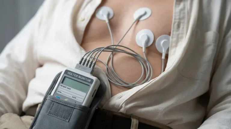
Health & Imaging
Abnormal EKG Test Results: What They Mean and What Happens Next

Quick answer: An abnormal EKG means the electrical pattern of your heartbeat fell outside the expected range on that recording. It does not automatically mean you have heart disease. Many abnormal findings are minor, common, or normal variants, especially in athletes and younger adults. Others, such as atrial fibrillation or signs of reduced blood flow, need prompt follow-up. Your doctor reads the EKG together with your symptoms, age, history and other tests before deciding what it means for you.
If you have chest pain, fainting, severe shortness of breath, or a racing heartbeat that won’t settle, call 911 now. Do not wait for a scheduled appointment.
This guide explains what abnormal ekg test results usually refer to, which findings are common and often harmless, which ones doctors take more seriously, and what tests typically come next. It is general information to help you understand your report and prepare questions. It is not a diagnosis.
Need to Schedule a Diagnostic Appointment?
Whether you need an MRI, ultrasound, X-ray, or another diagnostic service, Advantage MRI is here to help you take the next step in your healthcare journey.
Submit your appointment request online, and our team will contact you to confirm your visit.
Book Your Appointment →What Does “Abnormal EKG” Actually Mean?
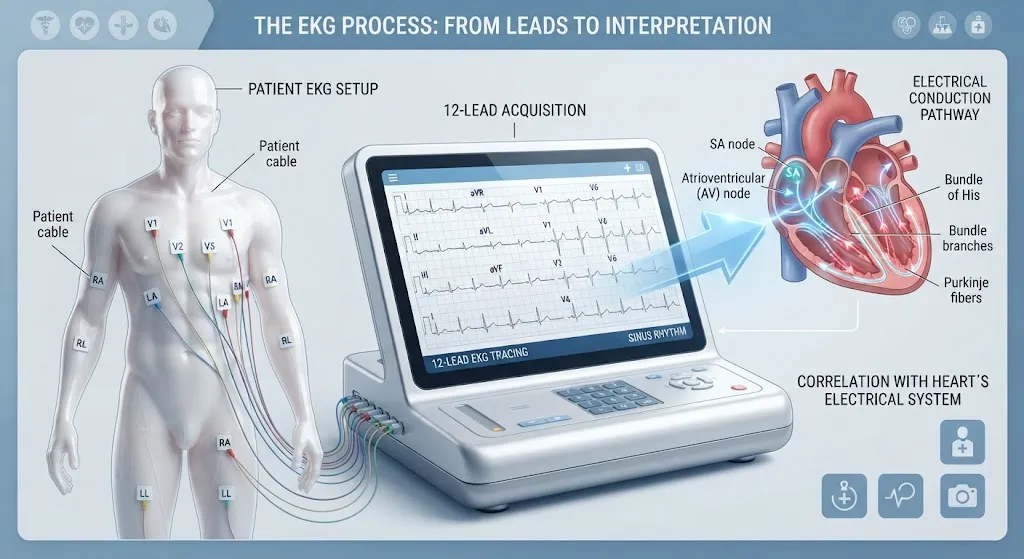
An electrocardiogram (EKG or ECG) records the electrical signals that make your heart beat. During a standard resting 12-lead EKG, ten small sticky electrodes are placed on your chest, arms and legs, and the machine records about 10 seconds of heart activity from 12 different angles. You can read a full overview in our guide to what an EKG test is.
The tracing is then measured against expected values:
- Heart rate (how fast)
- Rhythm (whether the beats are regular and start in the right place)
- Intervals (how long each part of the electrical cycle takes)
- Axis (the overall direction of electrical flow)
- Wave shape and size (P waves, QRS complexes, ST segments and T waves)
“Abnormal” simply means one or more of these measurements fell outside the normal range. That is a description of the tracing, not a verdict on your health.
Why the Machine Says “Abnormal” So Often
Most EKG machines print an automated interpretation at the top of the page, such as “Abnormal ECG” or “Borderline ECG.” These computer reads are designed to be cautious. They flag anything that could matter, which means they also flag harmless variants. A physician reviews the tracing and either confirms or overrides the computer’s statement. If you saw “abnormal” in a patient portal before talking to anyone, keep that in mind.
Normal EKG Ranges at a Glance
Your doctor will compare your results with ranges like these. Exact cut-offs vary slightly between references and labs.
| Measurement | Typical adult reference range | What it reflects |
|---|---|---|
| Heart rate | 60-100 beats per minute at rest | Speed of the heartbeat |
| PR interval | 120-200 milliseconds | Time for the signal to travel from the upper to the lower chambers |
| QRS duration | Under about 120 milliseconds | Time for the lower chambers (ventricles) to activate |
| QTc interval | Roughly under 450 ms (men) and 460 ms (women) | Time for the ventricles to activate and recover, corrected for heart rate |
| Rhythm | Regular “sinus rhythm” | Beats start in the heart’s natural pacemaker |
A value slightly outside one of these ranges is not the same as a dangerous result. Context matters a great deal.
Common Abnormal EKG Findings and What They Usually Mean
Below are findings people often see on an EKG report. The descriptions are general. Only a clinician who knows your history can tell you what a finding means for you.
Sinus Bradycardia (Slow Heart Rate)
A resting heart rate below 60 beats per minute. In fit people and athletes, this is often normal because a well-conditioned heart pumps more blood with each beat. It can also be caused by certain medications, such as beta blockers, or by thyroid problems or electrical conduction disease. Doctors pay more attention when it comes with dizziness, fatigue or fainting.
Sinus Tachycardia (Fast Heart Rate)
A resting heart rate above 100 beats per minute with a normal rhythm. Common causes include anxiety, caffeine, fever, dehydration, pain, anemia or an overactive thyroid. It is usually a sign of something else going on in the body rather than a heart problem on its own.
Sinus Arrhythmia
The heart rate speeds up slightly when you breathe in and slows when you breathe out. This is a normal finding, especially in children and young adults, even though some machines label it.
Premature Beats (PACs and PVCs)
Premature atrial contractions (PACs) and premature ventricular contractions (PVCs) are early “extra” beats. Many people feel them as a skipped beat or a flutter. Occasional premature beats are very common in healthy hearts. Doctors look more closely if they are frequent, come in runs, or appear with symptoms or known heart disease. A longer recording, such as a Holter monitor, is often used to count them. Our guide to Holter monitoring for palpitations and PVCs explains how that works.
Atrial Fibrillation (AFib)
An irregular, often fast rhythm in which the upper chambers quiver instead of contracting normally. AFib matters because it increases the risk of stroke and can weaken the heart over time if the rate stays high. It is one of the findings that usually leads to prompt follow-up, including blood tests, an echocardiogram and a discussion of stroke prevention.
Heart Block (AV Block)
The signal from the upper to the lower chambers is delayed or interrupted.
- First-degree AV block is a lengthened PR interval. It is often harmless and common in older adults and athletes.
- Second-degree and third-degree blocks are more significant because some or all signals fail to reach the ventricles. These can cause dizziness or fainting and may require treatment, sometimes a pacemaker.
Bundle Branch Block
The electrical signal travels down two “branches” to the left and right ventricles. If one branch is slow, the QRS complex widens.
- Right bundle branch block can appear in healthy people but may also relate to lung or heart conditions.
- Left bundle branch block is more often linked to underlying heart disease and usually prompts further testing, such as an echocardiogram.
Left Ventricular Hypertrophy (LVH) Pattern
Tall QRS voltages can suggest a thickened heart muscle, often from long-standing high blood pressure. An EKG is not very accurate for this. Thin people and athletes can show high voltages with a normal heart. An echocardiogram measures wall thickness directly.
ST-Segment and T-Wave Changes
The ST segment and T wave reflect how the ventricles recover after each beat. Changes here can be a sign of reduced blood flow to the heart muscle (ischemia), a past or current heart attack, inflammation, electrolyte imbalances or medication effects. They can also be “nonspecific,” meaning they don’t point to a clear cause. New ST changes with chest pain are treated as an emergency.
Prolonged QT Interval
A long QT can raise the risk of a dangerous rhythm. Causes include certain medications (some antibiotics, antidepressants and anti-nausea drugs), low potassium or magnesium, and inherited conditions. If this shows up, your doctor may review your medication list and check electrolytes.
Q Waves
Certain Q waves can suggest a previous heart attack, sometimes one that happened without obvious symptoms. They can also be positional or normal in some leads. Further imaging usually clarifies the picture.
Early Repolarization
A pattern of slight ST elevation seen commonly in young, healthy people and athletes. It is usually benign but is sometimes flagged by automated reads.
Abnormal vs Serious: How Doctors Sort Findings
It helps to think of abnormal EKG results in three broad groups. This is a simplification, but it reflects how clinicians often approach a report.
| Group | Examples | Typical approach |
|---|---|---|
| Often benign or normal variants | Sinus arrhythmia, mild bradycardia in athletes, early repolarization, isolated PACs | Usually no further testing unless symptoms are present |
| Needs evaluation | Frequent PVCs, LVH pattern, bundle branch block, nonspecific ST-T changes, first-degree AV block with symptoms | Follow-up testing such as echocardiogram, Holter or stress testing |
| Needs prompt or urgent care | New ST elevation or depression with symptoms, high-grade heart block, sustained fast arrhythmias, AFib with very fast rate | Same-day or emergency evaluation |
Can an EKG Be Abnormal When Your Heart Is Fine?
Yes. False positives are common for several reasons:
- Electrode placement. Electrodes placed a little too high or too low can change wave shapes.
- Movement or shivering. Muscle activity creates “noise” that can look like an abnormal rhythm.
- Body type. Chest shape, breast tissue and body size can affect voltages.
- Normal variants. Athletes, younger adults and some ethnic groups commonly show patterns that look unusual but are healthy.
- Automated interpretation. Computer reads tend to over-call findings.
The reverse is also true. A normal resting EKG does not rule out every heart condition. It captures only a few seconds. Problems that come and go, like intermittent arrhythmias, or problems that only appear during exertion, such as some coronary artery disease, can be missed. That is why your doctor may order more tests even when the EKG looks normal.

What Happens After an Abnormal EKG?
The next step depends on the finding, your symptoms and your risk factors. Common follow-ups include:
1. A Repeat EKG
Sometimes the simplest step is to repeat the test with careful electrode placement, especially if the first tracing had artifact or was borderline. A repeat EKG also shows whether a change is new or has been present for years. If you have old EKGs from another provider, bring copies. Comparison is one of the most useful tools a reader has.
2. Echocardiogram
An echocardiogram is an ultrasound of the heart. It shows the size of the chambers, the thickness of the walls, how well the heart pumps and how the valves work. It is the usual next step for LVH patterns, bundle branch blocks, AFib, Q waves and many murmurs. Our article on echocardiogram vs EKG explains how the two tests complement each other.
3. Holter or Extended Rhythm Monitoring
If the concern is an irregular rhythm or premature beats, a Holter monitor records every heartbeat for 24 hours or longer while you go about your routine. Read more in our complete guide: What Is a Holter Monitor?
4. Stress Testing
When ST-T changes raise concern about reduced blood flow, a stress test checks how the heart responds to exercise or medication. This may be a stress EKG or a stress echocardiogram. See our comparison of stress echocardiograms and stress EKGs.
5. Blood Tests
Electrolytes (potassium, magnesium, calcium), thyroid function and a complete blood count can explain some abnormal rhythms and QT changes. If a heart attack is suspected, a troponin blood test is done urgently.
6. Cardiology Referral
For significant or unexplained findings, your primary care doctor may refer you to a cardiologist, who can order advanced testing and discuss treatment.
Questions to Ask Your Doctor About an Abnormal EKG
Bring these to your follow-up appointment:
- What exactly was abnormal on my EKG?
- Is this finding new, or could it have been there for a long time?
- Could my medications, caffeine intake or a recent illness explain it?
- Do my symptoms fit with this finding?
- Do I need more tests, and which ones?
- Should I change my activity or exercise until we know more?
- Which symptoms should send me to the emergency room?
Symptoms That Need Urgent Attention
Get emergency care if an abnormal EKG comes with any of these:
- Chest pain, pressure or tightness, especially spreading to the arm, jaw or back
- Fainting or near-fainting
- Severe shortness of breath
- A very fast or very slow heartbeat with dizziness
- Sudden weakness, confusion or trouble speaking
These symptoms matter more than any line on a printout.
Abnormal EKG Results in Women
Women’s EKGs differ in a few ways. Women tend to have slightly longer QT intervals and a slightly faster resting heart rate. Heart attack symptoms in women can be less “classic,” such as fatigue, nausea, jaw pain or shortness of breath without much chest pain. Breast tissue and electrode placement under or around the breast can also affect the recording. If you are a woman with symptoms and an EKG that is labeled abnormal, make sure your follow-up considers these differences. Our guide on how long an EKG test takes covers what to expect during the test, including practical tips for women.
How to Prepare for a Repeat or Follow-Up EKG
A resting EKG needs very little preparation, but small steps improve accuracy:
- Avoid applying body lotion or oil to your chest on the day of the test.
- Wear a two-piece outfit so you only need to remove your top.
- Tell the technologist about any medications, including over-the-counter products and supplements.
- Try to rest quietly for a few minutes before the recording and avoid talking or moving during it.
- If you have an old EKG on file elsewhere, bring a copy or ask for it to be sent.
If you are paying out of pocket, our article on EKG test cost explains typical pricing factors with and without insurance.
EKG Testing at Advantage MRI in Chicago
Advantage MRI is a diagnostic imaging and testing center at 2008 N Pulaski Rd, Chicago, IL 60639. Alongside MRI, X-ray and ultrasound, we perform EKG testing, echocardiograms, Holter monitoring, EMG and nerve conduction studies in one location. That means if your doctor orders a follow-up test after an abnormal EKG, such as an echocardiogram or a Holter monitor, you may be able to have it done at the same facility.
Our team works with referring physicians so results go back to the clinician who ordered your test. You can learn more about our team and accreditation on our About page. We welcome insured, out-of-network and self-pay patients.
Frequently Asked Questions About Abnormal EKG Test Results
Is an abnormal EKG something to worry about?
Not always. Many abnormal EKG results are minor or are normal variants. Some findings, such as atrial fibrillation, high-grade heart block or new ST changes, need prompt follow-up. Your doctor will interpret the result alongside your symptoms and history.
Can anxiety cause an abnormal EKG?
Anxiety can raise your heart rate and cause sinus tachycardia. It can also make you tense or move during the recording, which creates artifact. Anxiety does not cause structural heart findings such as Q waves or bundle branch blocks.
Can an EKG detect a blocked artery?
A resting EKG can show signs of reduced blood flow or a past heart attack, but it often looks normal in people with stable coronary artery disease. Stress testing or other imaging is usually needed to look for blockages.
How accurate is the computer reading on an EKG?
Automated interpretations are useful for screening but often over-call findings. A physician review is the standard before any decision is made.
What does “borderline EKG” mean?
It usually means a measurement is slightly outside the normal range without a clear abnormal pattern. Borderline results often turn out to be insignificant, but your doctor may repeat the test or compare it with older tracings.
Can dehydration or low potassium cause an abnormal EKG?
Yes. Electrolyte imbalances, especially low potassium or magnesium, can change T waves and the QT interval and can trigger extra beats. A simple blood test can check for this.
How long does it take to get EKG results?
The recording itself takes a few minutes. A physician typically reviews the tracing and sends the report to your referring doctor. Ask your provider when to expect a follow-up call.
Conclusion
An abnormal EKG is a signal to look closer, not a diagnosis. Many findings are harmless variants or have simple explanations like medications, caffeine or electrolyte levels. Others point toward conditions that deserve follow-up, usually with an echocardiogram, Holter monitor, stress test or blood work. The most useful thing you can do is ask your doctor exactly what was abnormal, whether it fits your symptoms, and what the next step should be.
Need an EKG or follow-up heart test in Chicago? Book an appointment online or call Advantage MRI at (773) 227-9777. We are open Monday to Friday, 9:00 AM to 5:00 PM, and Saturday, 9:00 AM to 1:00 PM. For updates and patient tips, follow us on Facebook and Instagram.